Collaborations
Solving imaging challenges together.
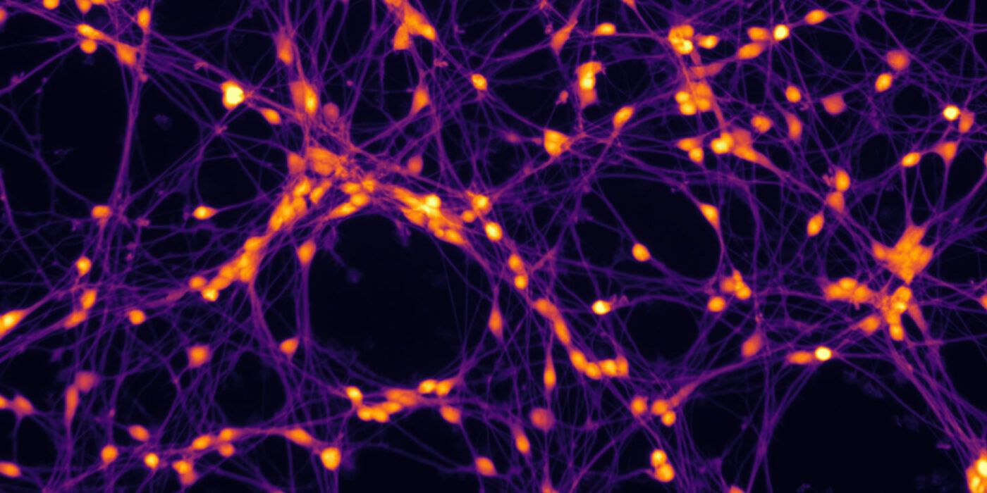
Image: J. Nyffeler, L. Reger, N. Teßendorf, UFZ | info
Collaboration Gallery

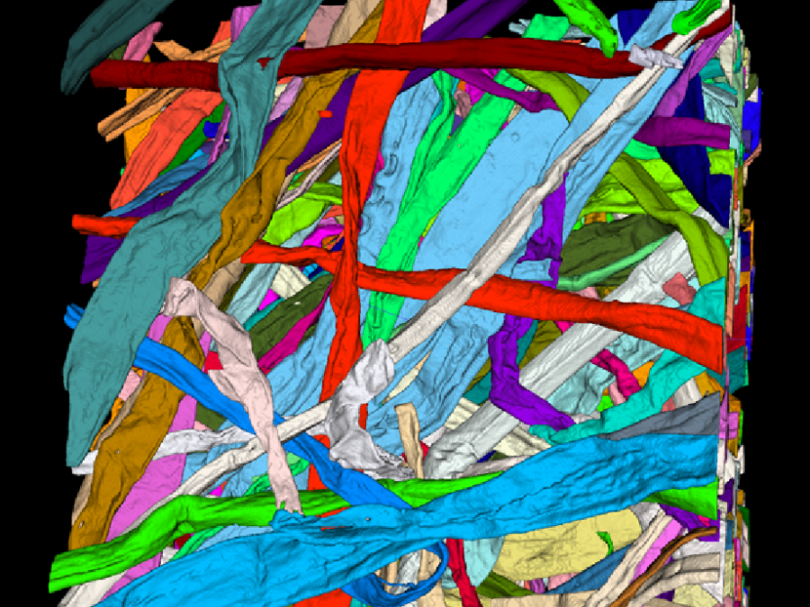
Active Learning-enabled Generation of (Patho-)Physiological Lung Architectures for Pulmonary Medicine (ALEGRA)
ALEGRA aims to discover new insights into the morphology, physiology, and functionality of mammalian lungs. Therefore, quantitative parameters will be extracted from light sheet fluorescence microscopy images featuring comprehensive annotations of unprecedented detail for structures of interest such as hollow airways, blood vessels, as well as alveoli. These annotations are made possible by a novel […]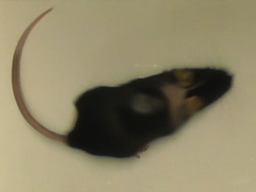
info
AI-based automatic analysis of mouse behavior in videos
The Helmholtz Imaging Engineering and Support Unit at DKFZ is developing an automated video analysis tool to better understand itch mechanisms and support the development of new treatments. By automatically detecting and quantifying scratching behavior in mice, the tool replaces time-consuming manual analysis and accelerates research.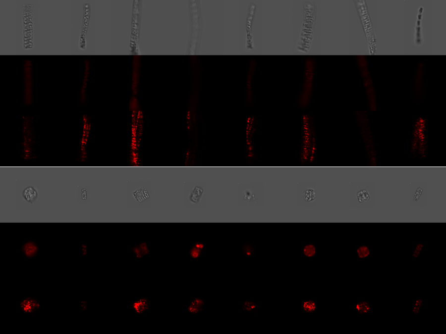
info
AI-based classification of phytoplankton community composition from field samples
Phytoplankton biodiversity is a key indicator of marine ecosystems health. Due to climate change derived impacts, phytoplankton communities worldwide are being affected by changes in the water temperatures and the access to nutrients and light. Current methods to quantitatively classify phytoplankton samples require taxonomy experts to manually count and classify the individual cells they see […]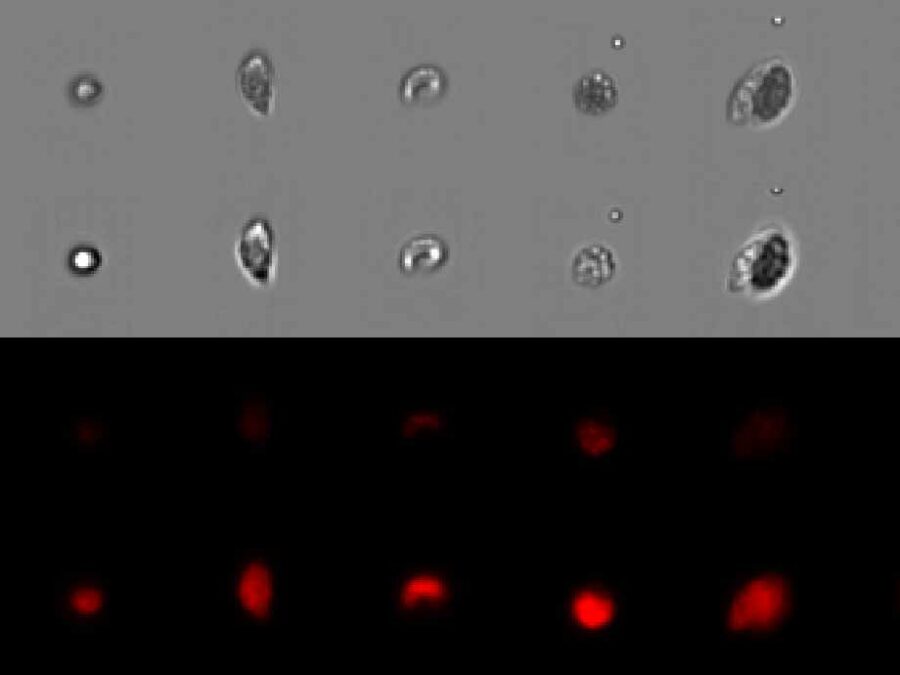
info
Automated Analysis of Evolutionary Experiments of Phytoplankton
There is a strong interest in understanding community assembly and dynamics. Experimental approaches using phytoplankton have proven to be extremely insightful to unravel underlying biological processes. Imaging flow cytometry is an emerging method becoming more and more popular in different fields. It allows us to capture changes within a community or population in a more […]
Image: A. Müller, D. Schmidt, M. Weigert, MDC | info
BetaSeg
Volume electron microscopy is the method of choice for the in situ interrogation of cellular ultrastructure at the nanometer scale, and with the increase in large raw image datasets generated, improving computational strategies for image segmentation and spatial analysis is necessary. Here we describe a practical and annotation-efficient pipeline for organelle-specific segmentation, spatial analysis and […]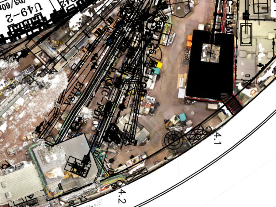
info
Capturing Reality at BESSY II
The experimental hall at BESSY II (Berliner Elektronenspeicherring) is a constantly evolving space, featuring a diverse range of structures, devices, and experiments managed by various departments, companies, and individuals. This dynamic nature necessitates regular updates to ensure the floor plan accurately reflects the hall’s current state. To address this, a video drone is deployed to […]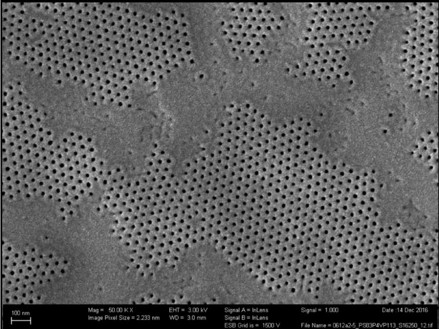
Connecting membrane pores and production parameters via machine learning (COMPUTING)
Isoporous block-copolymer membranes play a fundamental role in the filtration of liquids and can, for example, be used to purify drinking water. Despite recent progress in understanding the membrane formation process, finding suitable production parameters for a given precursor material (such as polymers of a certain length) still occurs in a trial-and-error fashion, wasting materials, […]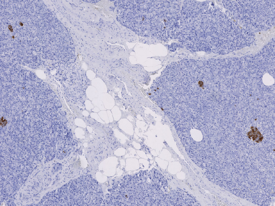
Detecting Type-2 Diabetes in histopathological images for a better understanding of biological processes behind the disease
Type-2 diabetes is a chronic disease affecting about 500 million people worldwide. Despite extensive research over the last decades, exact biological processes leading to a deteriorating insulin production are not yet fully understood. By building models that are able to classify whether a patient has type-2 diabetes or not from whole slide images of the […]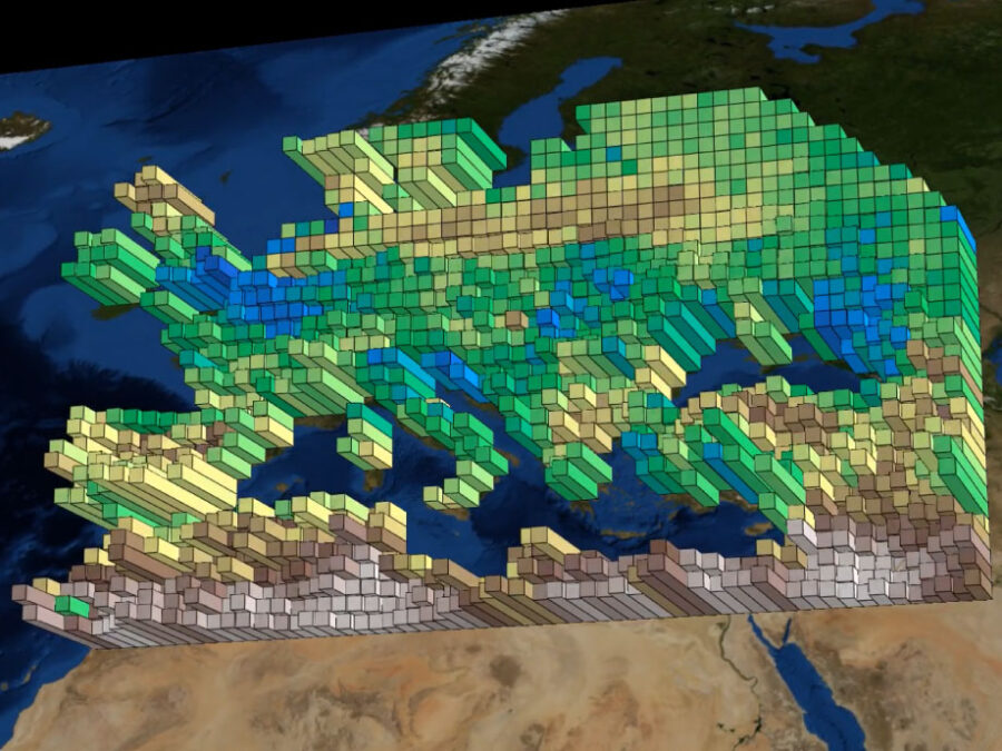
Image: Snapshots taken from videos created by the tool - original data by Almudena Garcia-Garcia and Jian Peng of Umweltforschungszentrum (UFZ) | info
EarthWatch
Earth-Watch introduces a Python package that visualizes spatiotemporal netCDF data on world maps by generating dynamic videos with PyVista. Designed to enhance the understanding and presentation of geospatial datasets over time, the tool allows users to overlay multiple observations, enabling comparative analyses and revealing correlations between different datasets. Additionally, it facilitates effective communication and presentations. By […]
Extracting clinically relevant parameters from real-time MRI images of fontan hearts
Obtaining accurate segmentations of the heart in real-time MRI allows a more realistic view on clinically relevant parameters, such as the stroke volume. Cardiac real-time MRI can assess diastolic filling under breath maneuvers or other cardiac load situations which potentially enhances diagnostics other than CINE breath hold cardiac MRI. Real-time MRI allows rapid acquisitions during […]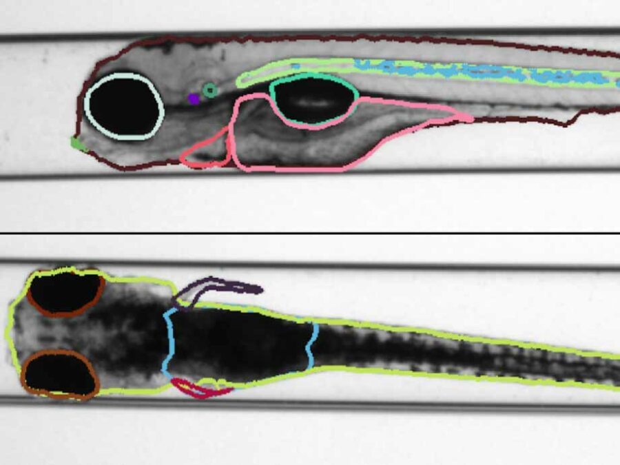
info
Identifying structural features of zebrafishes, using Semantic Segmentation
Zebrafish have a certain genetic similarity to humans and vertebrates. Particularly, the use of embryos is attractive due to the small scale screening capacity. Furthermore, similar to genuine cellular in vitro approaches, zebrafish embryos are considered as alternatives to animal testing. Therefore, they play a fundamental role in the detection of environmental and human health […]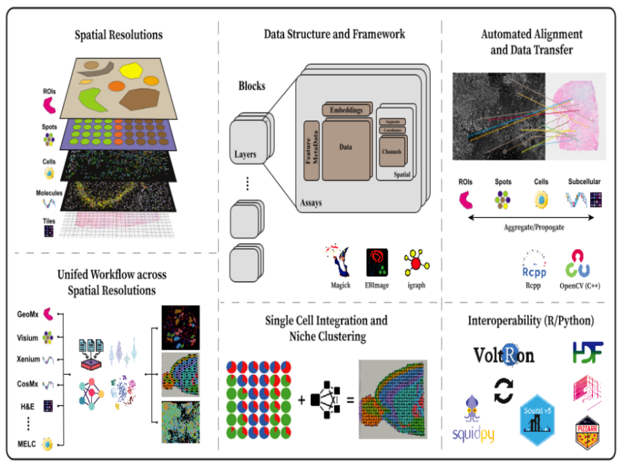
info
Image Registration for VolTron
VoltRon is an R based spatial omic analysis toolbox designed for multi-omics integration through spatial image registration. Focused on image registration, VoltRon leverages OpenCV – fully embedded into the package using Rcpp – to detect common features across images, ensuring precise alignment of multiple spatially-aware data modalities. The toolbox includes built-in mini Shiny apps that […]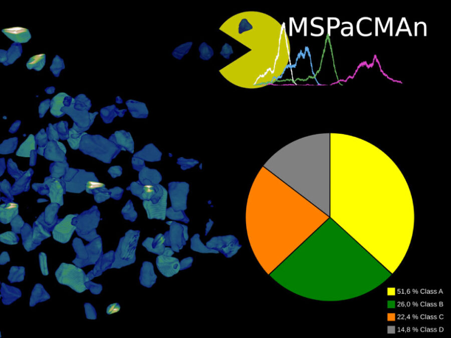
Image: Dr. Jose Ricardo da Assuncao Godinho, HZDR, Jan Philipp Albrecht, MDC | info
MSPaCMAn
MSPaCMAn is a workflow for quantifying mineral phases in 3D images of particulate materials using X-ray computed micro-tomography, designed to minimize imaging artifacts. It involves dispersing particles into samples, optimizing image processing to label individual particles, and identifying phases at the particle level by interpreting the grey-values of all voxels within each particle. This method […]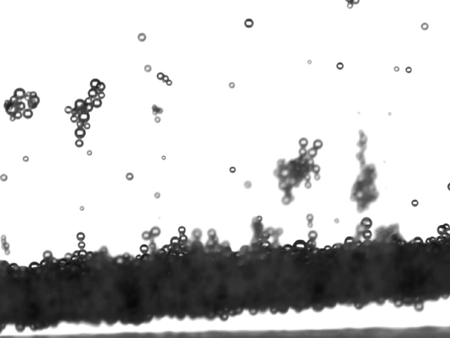
Optimizing electrode geometry and surface for hydrogen production
Electrolysis of water into oxygen and hydrogen is a cornerstone of modern energy storage, electric mobility and the transition towards a net-zero-emissions industry. Maximizing the efficiency of this technology is key to its economically viable wide scale adoption. One approach to both reduce the costs and improve the overall efficiency is to enhance the bubble […]
Paving the way for future mineral processing and recycling technologies through large-scale analysis of particulate samples
Understanding and quantifying the exact composition of mineral samples paves the way towards advanced methodologies that not only increase the effectiveness of ore processing but also enable future recycling technologies. To this end, samples consisting of ground particles embedded into an epoxy matrix are imaged with computed tomography (CT) at micrometer resolution. Due to the […]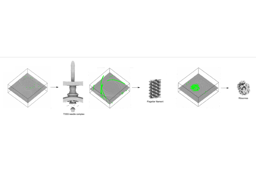
info
Pick Yolo
In collaboration with the Center for Structural Systems biology, Helmholtz Imaging has developed and trained a convolutional neural network for the picking of instances of proteins in cryoelectrontomograms (CryoET). The so picked instances are subsequently used to reconstruct a 3D structure of the proteins using subtomogram averaging methods. The novel picking methods exploit the well […]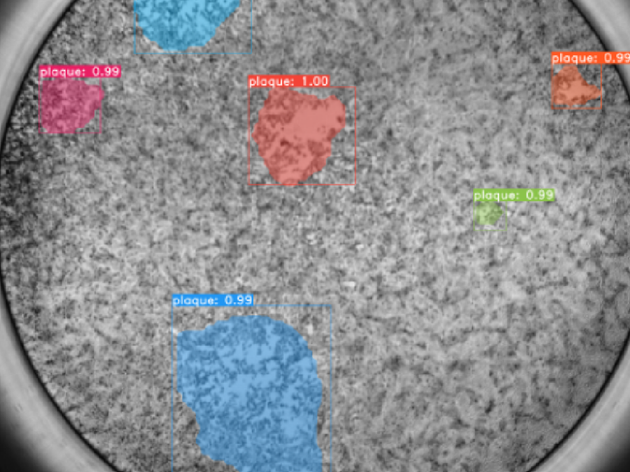
Plaque Assays
In collaboration with the Leibniz Institute of Virology and the Center for Structural Systems biology, Helmholtz Imaging is working on a processing pipeline for plaque assays. Plaque Assays are used to e.g. estimate the virus concentration in a given sample. For this high resolution images of cell cultures which have been infected with a virus […]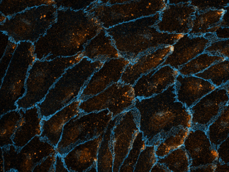
Image: Olya Oppenheim, MDC, Emir Bora Akmeriç, MDC, Jan Philipp Albrecht, MDC, Wolfgang Giese, MDC | info
Polarity-JaM
Cell polarity involves the asymmetric distribution of cellular components, cell shape, and contacts with neighboring cells. Gradients and mechanical forces bias cell polarity, coordinated by communication between adjacent cells. Advances in fluorescence microscopy and deep learning for image segmentation have expanded our understanding of cell polarity in health and disease. Polarity-JaM is an open-source package […]
Predicting Perovskite Thin-Film Photovoltaic Performance from Photoluminescence Videos
Photovoltaics are a key technology to decarbonize the generation of energy. While perovskite thin-films are a promising option to build powerful next generation photovoltaics demonstrating high power conversion efficiencies, their manufacturing process remains unstable. We build a model that directly predicts the solar cell performance based on a video capturing the perovskite layer formation prior […]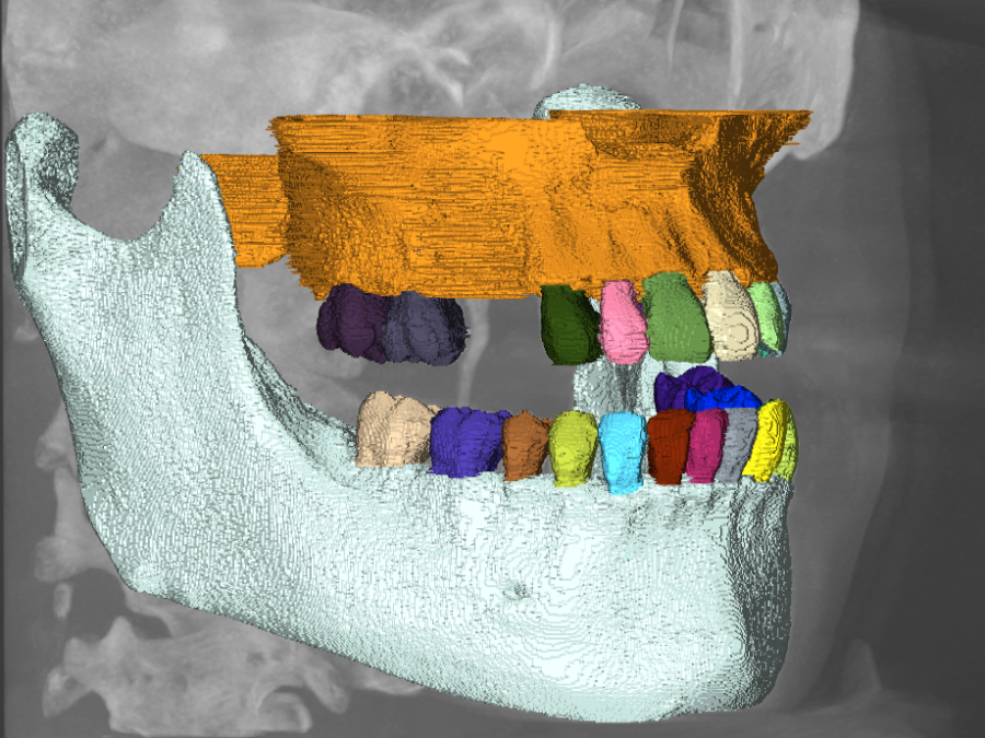
Segmentation and identification of tooth instances in cone-beam CT scans
The three dimensional labeling and identification of teeth in cone-beam CT scans is a time-consuming and challenging process which is exacerbated by (partially) missing teeth, tooth misalignments as well as the presence of implants or other utilities like metal plates or wires. As part of this project we develop an AI-based instance segmentation algorithm to […]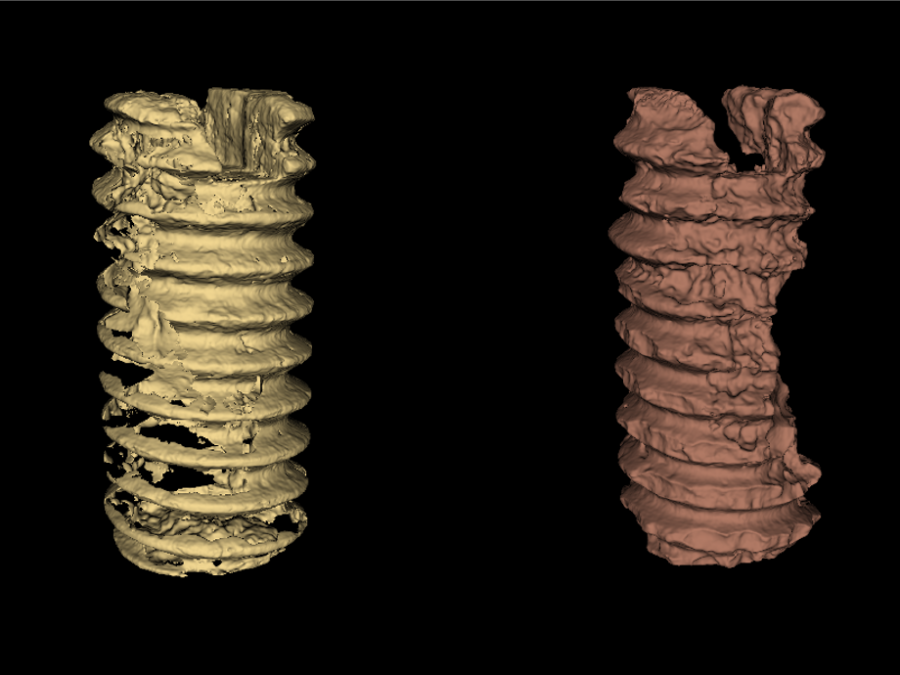
Segmentation of biodegradable bone implants
Together with the experts from Hereon the Helmholtz Imaging Support Team collaborates on segmenting synchrotron CT data. In 2021 the collaboration on semantic segmentation of biodegradable bone implants using a U-net resulted in two publications acknowledging the contribution of Helmholtz Imaging [1],[2]. Beyond that, we studied “Instance segmentation of paper fibers” imaged at the Hereon […]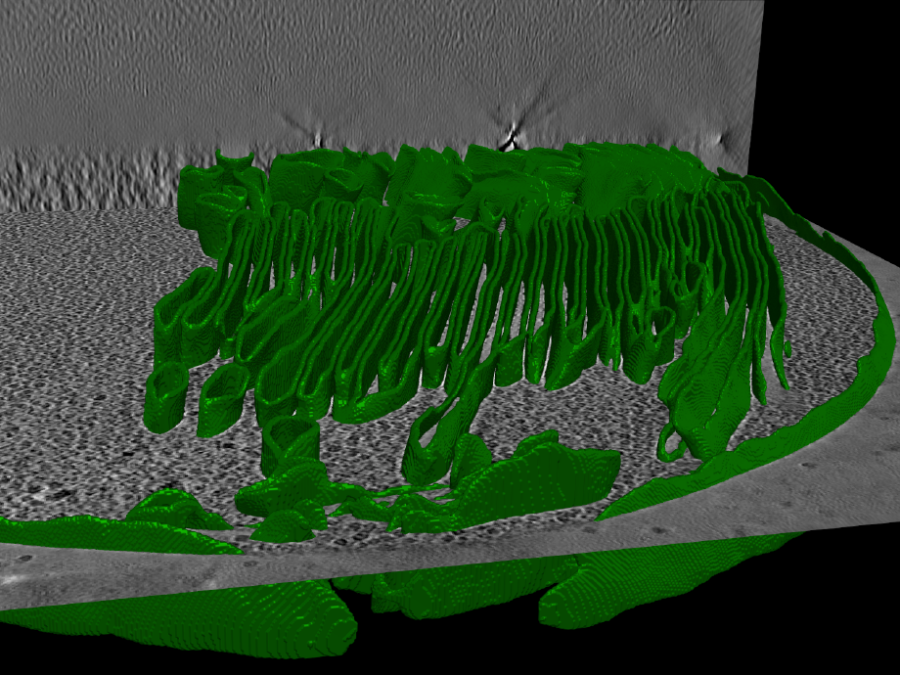
Segmenting cell membranes in cryoET data
In this project we develop semantic segmentation methods for accurate delineation of cell membranes in cryoET data. Not only do these segmentations enable the analysis of the spatial configuration of membranes, for example in chloroplasts, they also facilitate downstream analysis like determining the distribution of membrane proteins on their surface.
Single hit detection in single particle imaging with XFELs
Single particle imaging with x-ray-free electron lasers enables unique insights into the inner structure of nanometer-sized biological particles such as viruses. In order to reconstruct their 3D composition, a large number of 2D diffraction patterns must be acquired. AI-based isolation of single hits from among the hundreds of thousands of acquisitions made throughout the course […]
Ultra Content Screening for Clinical Diagnostics and Deep Phenotyping
For the Helmholtz Imaging project UCS (Ultra Content Screening for Clinical Diagnostics and Deep Phenotyping) we developed software to allow the project partners to process their immunofluorescence labelled light microscopy data of nuclei and cells for single cell proteomics. The developed pipeline includes instance segmentation of nuclei and cells in the immunofluorescence assays, registration of […]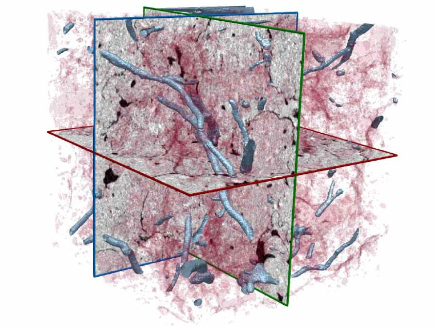
info
Understanding and analyzing biopores and plant roots in Soil Cores using Semantic Segmentation of CT Images
To understand how processes in ecosystems work and how they are connected the analysis of soil systems is essential. Since traditional computer vision methods for analysing soil cores reach their limits the next step is to integrate deep learning methods. Therefore a sufficient amount of labeled ground truth data is needed. Since labeling this large […]
Understanding and analyzing plant roots using semantic segmentation of MRI images
The optimization of plants has long focused on the above-ground parts. Recently, new efforts are being made to exploit the potential below ground. To this end, our partners at the FZJ have developed an imaging system which enables imaging the root system throughout the growth of the plants using MRI. Besides qualitative analysis, a precise […]
Understanding lung diseases and optimizing their treatment
Understanding the distribution of nanoparticles after inhalation can lead to the discovery of novel, more effective drug delivering techniques. To this end we develop AI-based methods for semantic segmentation of the airways in non-dissected whole murine lungs, imaged with light sheet fluorescence microscopy. The analysis of airway properties in diseased lungs will furthermore shed light […]