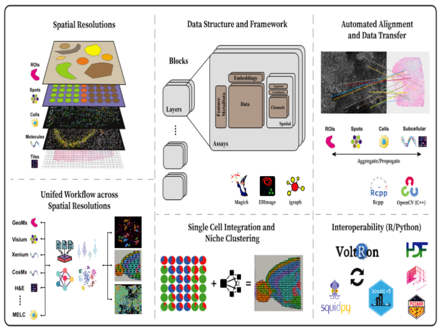
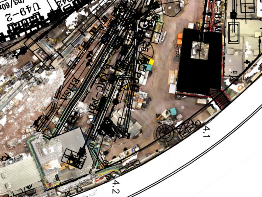
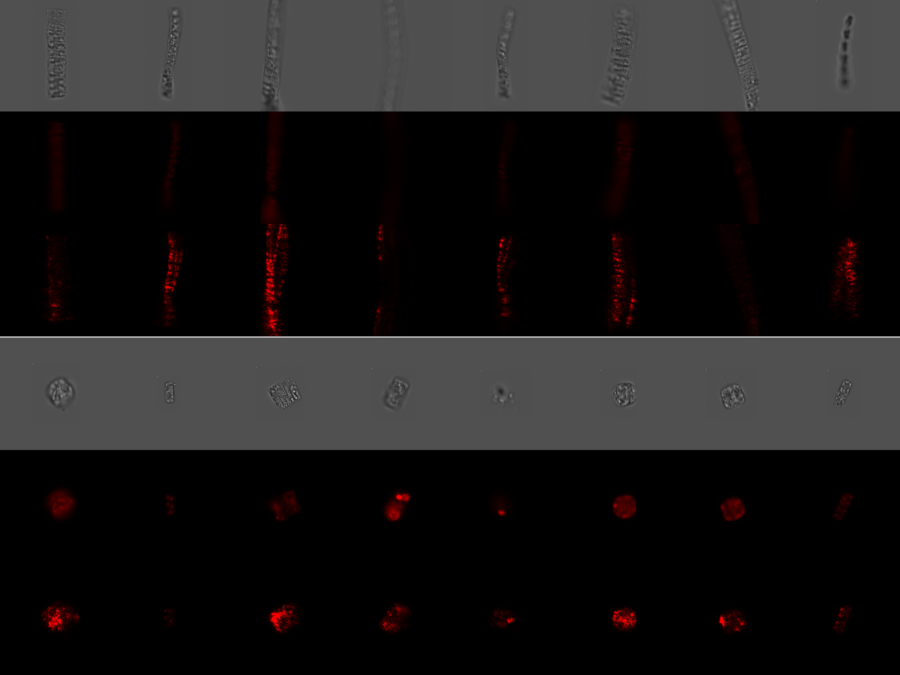
Helmholtz Imaging Collaborations are your opportunity to directly tap into the expertise of Helmholtz Imaging. As part of a Collaboration, you will team up with one or multiple of our units and closely work together on solving imaging-related problems. A Collaboration is of collaborative nature and necessitates the active contribution of all parties involved. Collaborations can run for up to six months or longer, allowing us to take a deep dive into a specific problem and solve it for good!
Suitable topics for a collaboration are anything that is related to imaging and image analysis. Our Helmholtz Imaging Units offer a wide range of expertise covering the entire imaging pipeline, from the development of new imaging modalities all the way to AI-based image analysis. Whether you require support in (AI-based) image analysis, image reconstruction, annotation, data management, software solutions, addressing inverse problems, developing novel imaging modalities, 3D visualization and more – we are your partner. For more information about the expertise of our units, take a look at our team page.
Interested in a Collaboration? Apply now!
The application process is simple and straightforward: Applications are handled via the Support Hub. Either send us an email or use our application form.
Your application should include the following information:
- Problem statement,
- Desired outcome / how can HI help solve the problem,
- List of participating researchers,
- Contact information of main applicant,
- Depending on the task additional information is considered beneficial (e.g. sample data, respective annotations, prior publications etc.),
- (optional) If you have already been in contact with one of our units or this application has emerged from an interaction initiated by the Support Hub feel free to name a desired Helmholtz Imaging unit to collaborate with
Review of your application: We will then review your application and, unless a desired partner unit is specified, match you with one of our units. If your application is accepted, you and the designated Helmholtz Imaging unit will then enter the evaluation phase.
Evaluation phase (duration 1-2 weeks): The goal of the exploration phase is to assess the feasibility of the project, agree on a desired (quantifiable) outcome and set a preliminary timeline.
Working Phase (up to 6 months): Following a successful evaluation phase, you and your Helmholtz Imaging collaborators work on the actual project.
If the time is up and the predefined goals are not yet reached, both partners assess the current state of the project and evaluate whether a prolongation is desired and may lead to the desired outcome. If both parties agree, the Collaboration can be extended.
Just like the rest of our portfolio, Helmholtz Imaging Collaborations are free of charge. Helmholtz Imaging Collaborations do not offer opportunities for obtaining additional funding. Any contribution of Helmholtz Imaging is subject to our Publication Policy.


