Single hit detection in single particle imaging with XFELs
Single particle imaging with x-ray-free electron lasers enables unique insights into the inner structure of nanometer-sized biological particles such as viruses. In order to reconstruct their 3D composition, a large number of 2D diffraction patterns must be acquired. AI-based isolation of single hits from among the hundreds of thousands of acquisitions made throughout the course of a single experiment is quintessential for the success of the reconstruction.
Publication:
Assalauova, Dameli, et al. “Classification of diffraction patterns using a convolutional neural network in single-particle-imaging experiments performed at X-ray free-electron lasers.” Journal of Applied Crystallography 55.3 (2022).
Source code:
https://gitlab.hzdr.de/hi-dkfz/applied-computer-vision-lab/collaborations/desy_2021_singleparticleimaging_cnn
Dataset:
https://zenodo.org/record/6451444
Other Collaborations

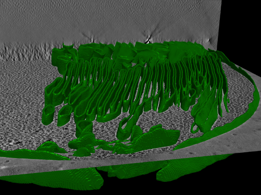
Extracting clinically relevant parameters from real-time MRI images of fontan hearts
Obtaining accurate segmentations of the heart in real-time MRI allows a more realistic view on clinically relevant parameters, such as the stroke volume. Cardiac real-time MRI can assess diastolic filling under breath maneuvers or other cardiac load situations which potentially enhances diagnostics other than CINE breath hold cardiac MRI. Real-time MRI allows rapid acquisitions during […]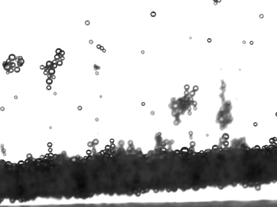
Optimizing electrode geometry and surface for hydrogen production
Electrolysis of water into oxygen and hydrogen is a cornerstone of modern energy storage, electric mobility and the transition towards a net-zero-emissions industry. Maximizing the efficiency of this technology is key to its economically viable wide scale adoption. One approach to both reduce the costs and improve the overall efficiency is to enhance the bubble […]