Helmholtz Imaging Projects
Helmholtz Imaging Projects aim to initiate cross-cutting research collaborations and identify innovative research topics in the field of imaging and data science.
Funds for Helmholtz Imaging Projects are annually granted to cross-disciplinary research teams for collaborative mid-term projects.
Ideally, Helmholtz Imaging Projects are co-created with users and non-academic stakeholders to ensure the quick adoption of results.
Funding for the first Helmholtz Imaging projects started in December 2020. Many teams have since begun work on major challenges and pressing issues facing society to develop sustainable solutions for tomorrow and beyond.
Discover these outstanding and fascinating research projects with us or become a part of Helmholtz Imaging Projects and apply for your own project. The next call for Helmholtz Imaging Projects is OPEN until July 30, 2025. Find out more about the project call in this summary.
Helmholtz Imaging Projects – completed
Avanti
X-ray tomoscopy of dynamic manufacturing processes
How can the manufacturing processes of materials be mapped at the smallest level? How do you train an artificial intelligence to analyze these processes automatically? That’s the focus of the Avanti project, which aims to improve X-ray tomoscopy – the imaging and quantification of three-dimensional images of very fast-moving processes.
Deep4OM
Deep learning powered optoacoustic mesoscopy for non-invasive diagnostics of skin diseases
Deep4OM aims to develop a deep learning-based framework for optoacoustic mesoscopy image analysis, enabling quantification of human skin biomarkers for non-invasive skin disease diagnosis. Deep4OM has the potential to change the landscape of non-invasive skin imaging, and could significantly promote the diagnostic and prognostic applications of RSOM in clinical routine.
HighLine
High Image Quality for Lines in MRI: From Roots to Angiograms
MR images of roots and vessels are very similar: both display thin, line-like objects. The aim of the project is to increase image quality of both kind of MR data by exploiting their similarity. HighLine aims at obtaining high quality images in reduced scan time to lower patient burden and increase patient and plant throughput by adapting state-of-the-art 3D image enhancement methods, and developing new deep-learning based methods.

Hyper 3D-AI
Artificial Intelligence for 3D multimodal point cloud classification
The aim is to develop an artificial intelligence that can achieve the fusion of two-dimensional data with three-dimensional information. Based on this, the software would simultaneously be able to recognise image characteristics as well as the spatial relationships between different objects.

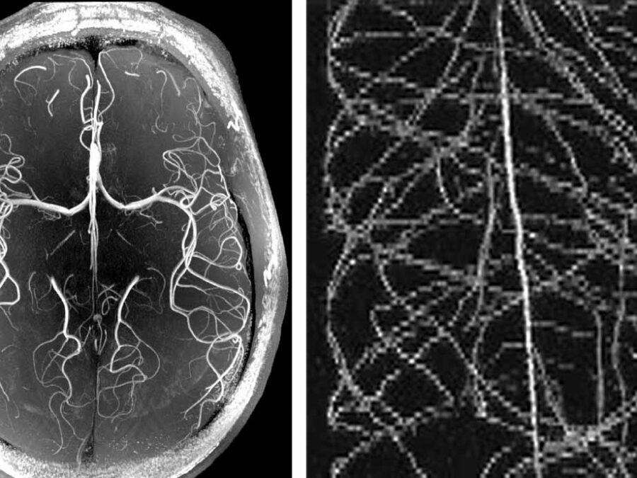
JIMM
Geophysical Joint Inversion for Accurate Brain Myelin Mapping
The aim of this project is to develop a method for clinically diagnosing neurodegenerative diseases. The content of myelin in the brain – a substance that becomes degraded in diseases – will be quantified using methods from geophysics in order to facilitate early detection and treatment.

MultiSaT4SLOWS
Multi-Satellite imaging for Space-based Landslide Occurrence and Warning Service
In order to detect impending landslides before they occur and to enable reliable emergency mapping after a landslide, the researchers are combining optical data with radar data from satellites. Using machine learning methods, computers will be trained to recognise the tiniest of changes in things like sloping landscape surfaces.

NImRLS
Neuroimaging Biomarkers for Restless Leg Syndrome
The aim is to develop a software solution that can analyse enormous amounts of data on tens of thousands of subjects from large-scale health studies. Using restless leg syndrome as an example, genomic data will be combined with neuroimaging data in order to identify new biomarkers with the help of machine learning methods.

SATOMI
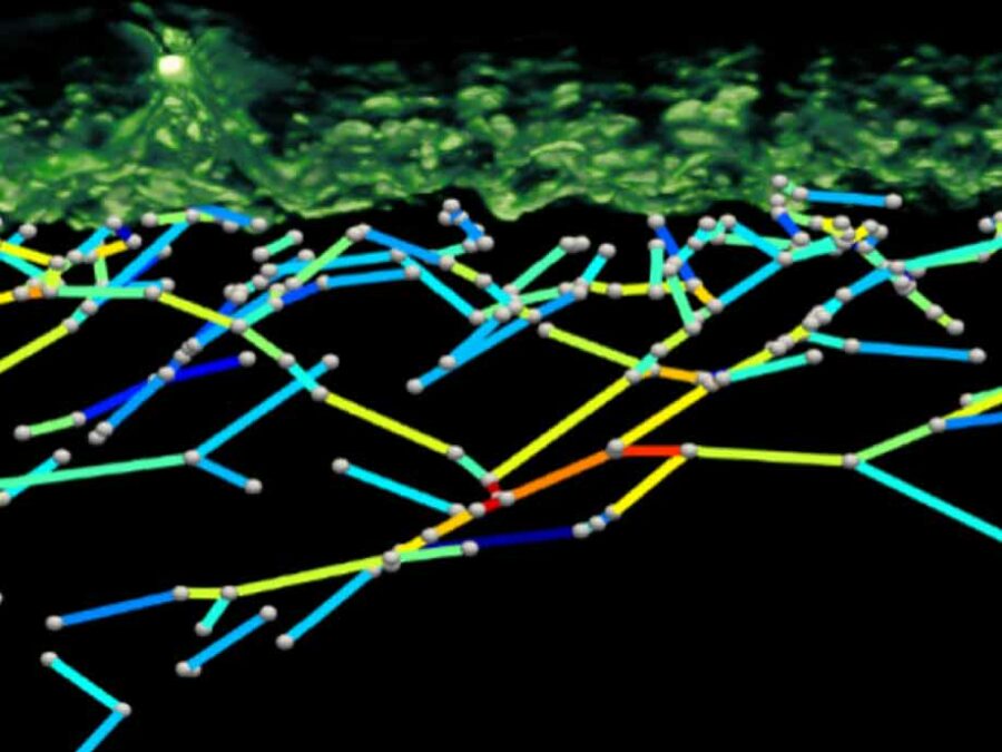
Tackling the segmentation and tracking challenges of growing colonies and microbialdiversity
An artificial intelligence will observe the growth of bacteria: from microscope images of bacterial cultures taken at regular intervals, it will precisely track the development and division of individual cells – even when multiple bacterial species are cultivated together.
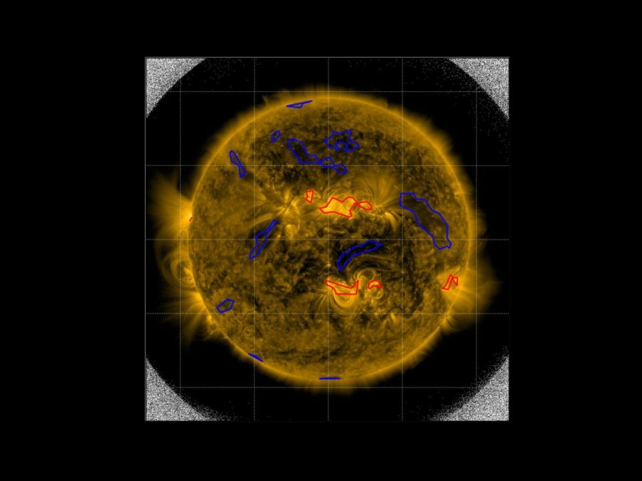
SIM
Solar Image-based Modelling
The aim of the project is to develop an algorithm by which computers can automatically predict the space weather. This will make use of datasets of solar images that have been captured from space. The method could replace computationally demanding physics-based models and deliver space weather forecasts long before the effects of solar events are […]

SmartPhase
Fast, intelligent 3-D X-ray images for material examination
In order to be able to improve materials, it helps to take a look at their microstructures. This is because valuable information about their properties and behaviour can be found there – for example information about when which ageing processes begin. The aim of this project is to automate and accelerate access to this information with the help of a smart imaging technique.

UCS
Ultra Content Screening for Clinical Diagnostics and Deep Phenotyping
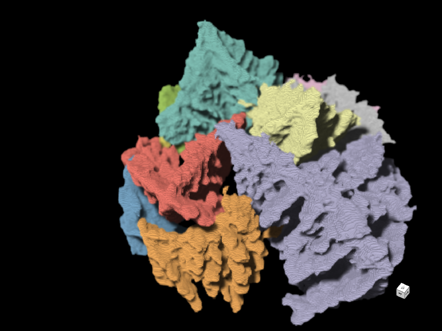
A method will be developed in which selected biomarkers in tumour and bone marrow cells from cancer patients will be examined and analysed automatically. The novel technology is based on ultra content screening technology, which allows detailed insights at the single cell level.

UTILE
Autonomous image analysis to accelerate energy materials discovery and integration
Research into green materials for clean energy generation is moving at full speed – yet still requires a long time to complete. This project is working on an open source image processing application that uses artificial intelligence to drive the analysis and management of image data from experiments across the energy materials community.