Deep4OM
Deep learning powered optoacoustic mesoscopy for non-invasive diagnostics of skin diseases
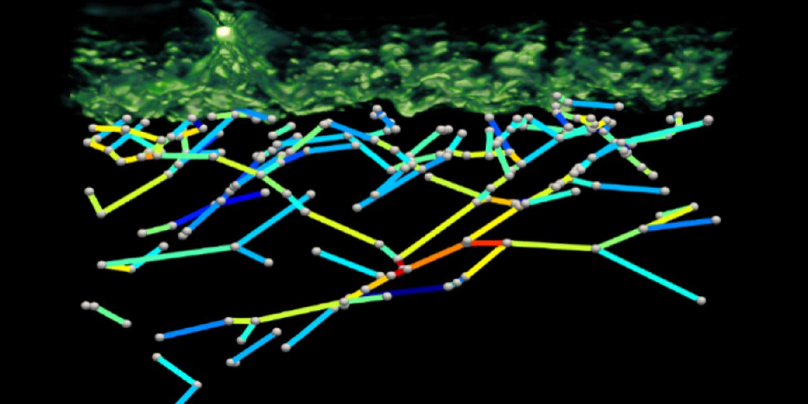
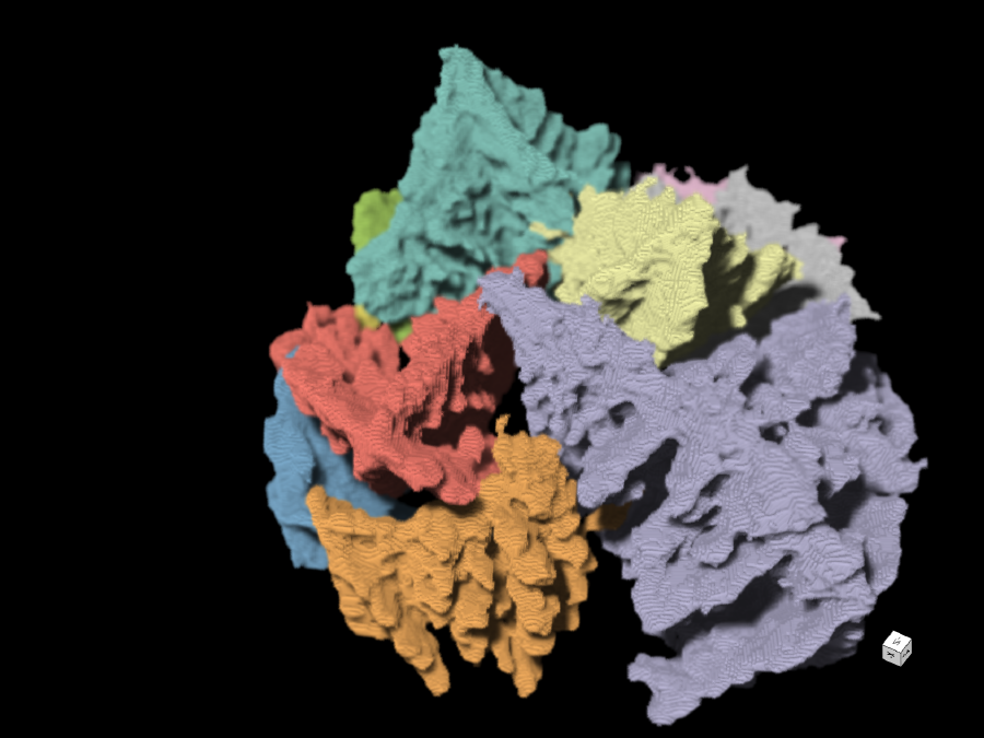
Raster-scan optoacoustic mesoscopy (RSOM) has achieved unprecedented resolutions in the non-invasive assessment of human skin anatomy and vasculature in vivo through the whole skin depth. It shows great potential for diagnostics and therapy monitoring in skin and other diseases. However, automated and quantitative analysis of three-dimensional RSOM datasets remains a challenge and prevents its clinical translation.
Deep4OM aims to develop the first quantitative image analysis framework based on deep learning techniques that is able to automatically analyze the high resolution RSOM images and derive various skin biomarkers for disease classification. The developed method will be tested on RSOM and histological data acquired from patients suffering from various skin diseases. As such, Deep4OM has the potential to change the landscape of non-invasive skin imaging, and could significantly promote the diagnostic and prognostic applications of RSOM in clinical routine.
Other projects
Avanti
X-ray tomoscopy of dynamic manufacturing processes
How can the manufacturing processes of materials be mapped at the smallest level? How do you train an artificial intelligence to analyze these processes automatically? That's the focus of the Avanti project, which aims to improve X-ray tomoscopy – the imaging and quantification of three-dimensional images of very fast-moving processes.
JIMM
Geophysical Joint Inversion for Accurate Brain Myelin Mapping
The aim of this project is to develop a method for clinically diagnosing neurodegenerative diseases. The content of myelin in the brain – a substance that becomes degraded in diseases – will be quantified using methods from geophysics in order to facilitate early detection and treatment.
SmartPhase
Fast, intelligent 3-D X-ray images for material examination
In order to be able to improve materials, it helps to take a look at their microstructures. This is because valuable information about their properties and behaviour can be found there – for example information about when which ageing processes begin. The aim of this project is to automate and accelerate access to this information with the help of a smart imaging technique.