JIMM
Geophysical Joint Inversion for Accurate Brain Myelin Mapping

The research community has long been on the trail of a substance that could be a key to understanding many neurological diseases: myelin is a biomembrane that surrounds most neurons in the human brain. “In many diseases, like multiple sclerosis for example, the myelin content of the brain decreases,” explains Prof. Dr. Tony Stöcker of the German Center for Neurodegenerative Diseases (DZNE). “Myelin can therefore serve as a marker for early detection: if we observe in good time that the myelin content is decreasing, then we could catch the onset of a disease at an early stage.”
The trouble is that it has been difficult so far to precisely quantify how much myelin is present. Experienced radiologists can assess brain tissue subjectively from MRI scans, where the variations in contrast allow them to draw conclusions about the state of the myelin, however they cannot determine the exact amount.
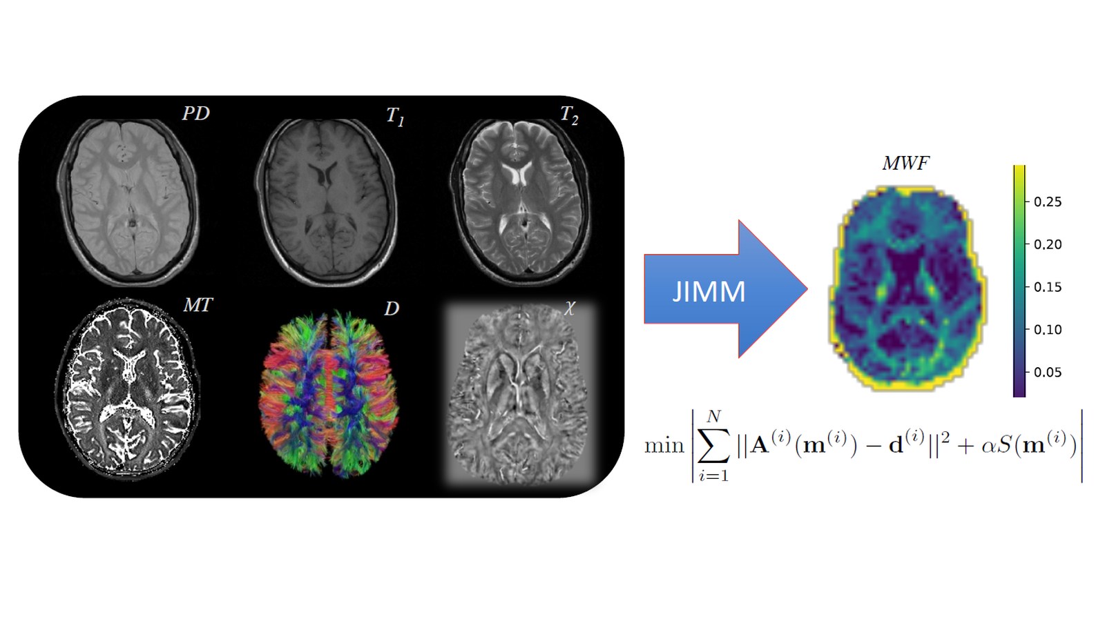
In this project, a method will be developed that can automatically calculate how much myelin is present in the brain from a combination of different MRI scans. The researchers will be using methods that are already being used in geophysics to deduce information about underground layers of earth from images of the ground surface. This involves complex mathematical tools and inversion methods, which have never before been used in medical imaging.
If successful, the new method could greatly facilitate the early detection and even the treatment of neurodegenerative diseases.
Other projects

SATOMI
Tackling the segmentation and tracking challenges of growing colonies and microbialdiversity
An artificial intelligence will observe the growth of bacteria: from microscope images of bacterial cultures taken at regular intervals, it will precisely track the development and division of individual cells – even when multiple bacterial species are cultivated together.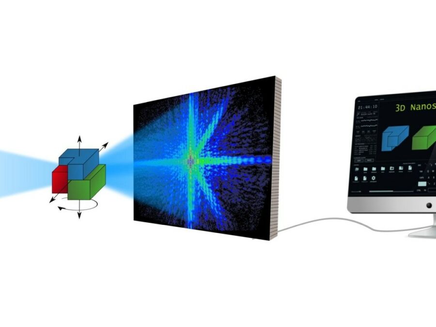
AsoftXm
Advanced Soft-X-Ray Microscopy Solutions
The project aims to develop a method that will speed up the analysis of diffraction patterns that arise in UV and soft X-ray light microscopy, so that the structure of the studied sample can be calculated more efficiently. The method could make the three-dimensional study of nanomaterials considerably easier. There are times when researchers need […]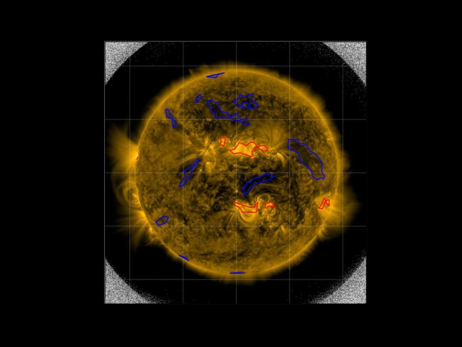
SIM
Solar Image-based Modelling
The aim of the project is to develop an algorithm by which computers can automatically predict the space weather. This will make use of datasets of solar images that have been captured from space. The method could replace computationally demanding physics-based models and deliver space weather forecasts long before the effects of solar events are […]