SATOMI
Tackling the segmentation and tracking challenges of growing colonies and microbialdiversity

PhD biology students in the past would have to make the best of their monotonous lab tasks: “We would hold regular cell-counting parties,” relates Prof. Dr. Dietrich Kohlheyer of Forschungszentrum Jülich. “We had beer and pizza, and at recurring intervals we would look through the microscope to count how the bacteria were multiplying over time.” However, when it comes to analysing more complex cell structures, counting is no longer an option.
This is where the research project comes in, which is aimed at developing a deep-learning method for analysing images from microscopes. “We will then use this method to watch bacteria as they grow,” Kohlheyer says. As the bacterial cultures grow under the microscope, pictures will be taken at set intervals. The task goes beyond simple counting: “We also want to know how the individual cells behave. So, for example, what daughter cells came about from the division of a parent cell?” says Dr. Katharina Nöh. She and her workgroup at FZ Jülich are developing software that can be used to track a cell family tree of sorts from images chock full of bacteria. The method of choice is called “probabilistic multi-object-tracking”, and the data processing for this method is being developed together with Prof. Ralf Mikut of KIT and Dr. Hanno Scharr of FZ Jülich. It is intended to work for all kinds of morphologies. Different bacteria namely develop different structures as they grow: some have daughter cells that split off, some have vesicles that burst open, while others grow out long like the branch of a tree. The aim is to develop artificial intelligence that can follow all of these variants, and even distinguish between individual cells among several bacterial species living and growing together.
SATOMI has secured follow-up funding. The project is now continuing under the name EMSIG (Event-driven Microscopy for Smart Microfluidic Single-cell Analysis).
Other projects
BENIGN
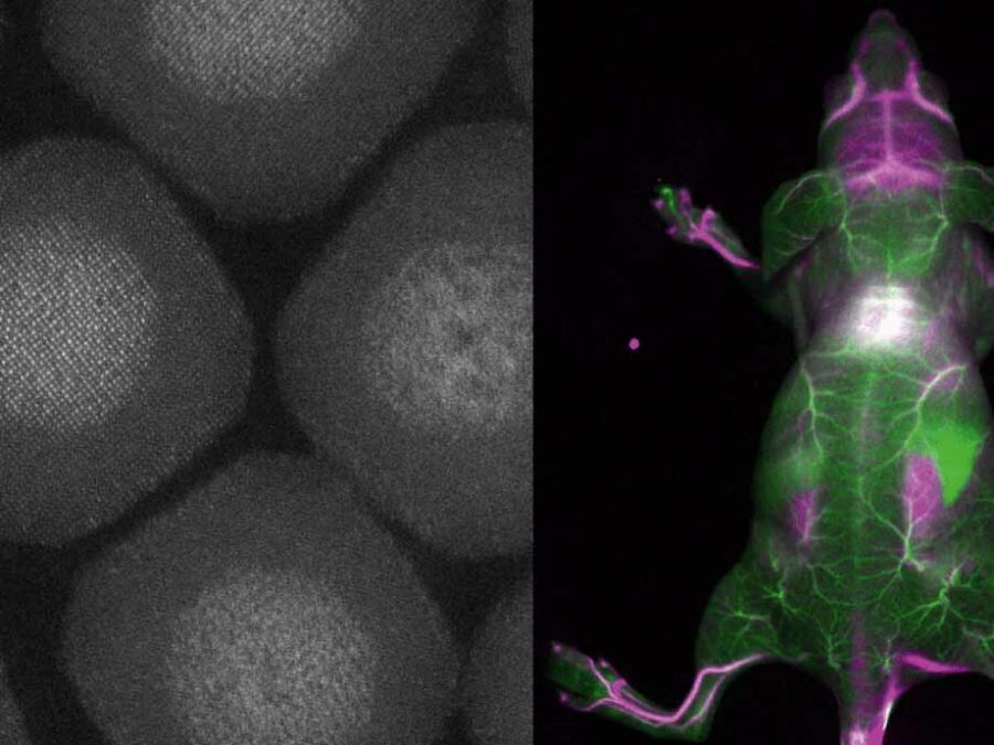
Biocompatible and Efficient Nanocrystals for Shortwave Infrared Imaging
The BENIGN project aims to enable non-invasive molecular imaging with cellular resolution in vivo at depths of several millimeters. This will be achieved using light from the shortwave infrared (SWIR) range (1000-2000 nm), which has less scattering and autofluorescence compared to the visible and near-infrared spectral range. Bright and targeted imaging agents are needed to fully exploit this range. The project will develop a new approach using lanthanide-based core-shell structures that emit light in the 1500-2000 nm range.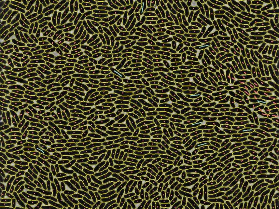
EMSIG
Event-driven Microscopy for Smart Microfluidic Single-cell Analysis
Microfluidic live-cell imaging (MLCI) unlocks spatio-temporal insights into population heterogeneity emerging from a single cell. EMSIG brings smart live-event detection capabilities to MLCI to facilitate the adaptive optimization of biological event resolution and autonomously counteracting deteriorating image qualities.
JIMM
Geophysical Joint Inversion for Accurate Brain Myelin Mapping
The aim of this project is to develop a method for clinically diagnosing neurodegenerative diseases. The content of myelin in the brain – a substance that becomes degraded in diseases – will be quantified using methods from geophysics in order to facilitate early detection and treatment.