Collaborations — Ongoing
Image Registration for VolTron
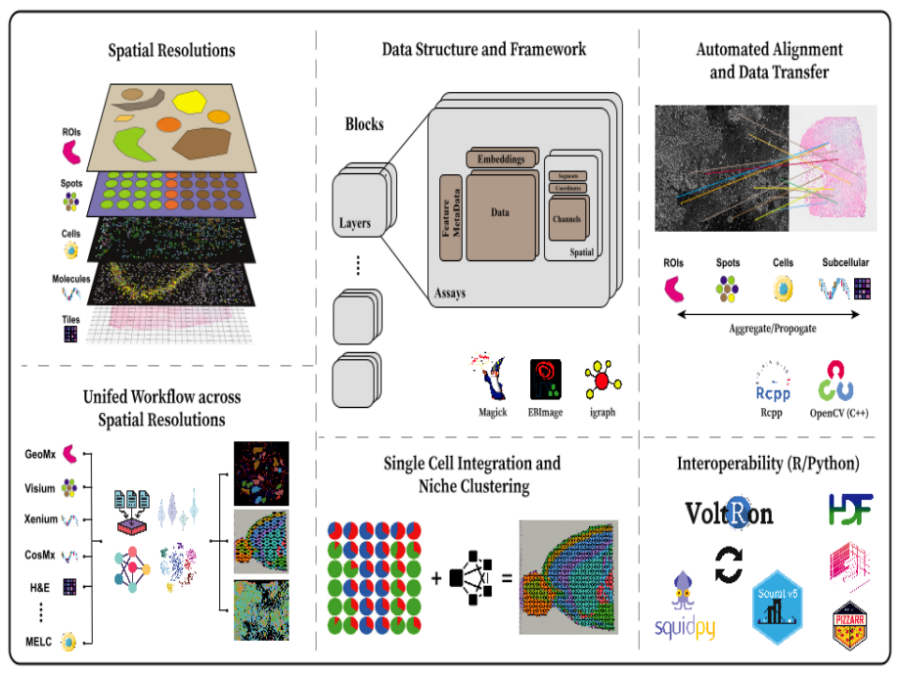
VoltRon is an R based spatial omic analysis toolbox designed for multi-omics integration through spatial image registration. Focused on image registration, VoltRon leverages OpenCV – fully embedded into the package using Rcpp – to detect common features across images, ensuring precise alignment of multiple spatially-aware data modalities. The toolbox includes built-in mini Shiny apps that […]
Capturing Reality at BESSY II
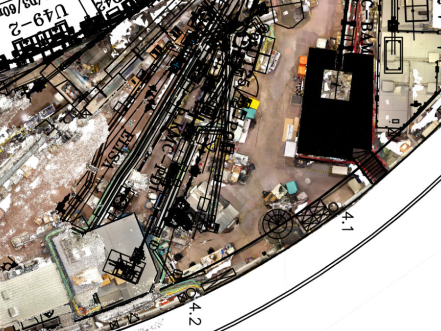
The experimental hall at BESSY II (Berliner Elektronenspeicherring) is a constantly evolving space, featuring a diverse range of structures, devices, and experiments managed by various departments, companies, and individuals. This dynamic nature necessitates regular updates to ensure the floor plan accurately reflects the hall’s current state. To address this, a video drone is deployed to […]
AI-based classification of phytoplankton community composition from field samples
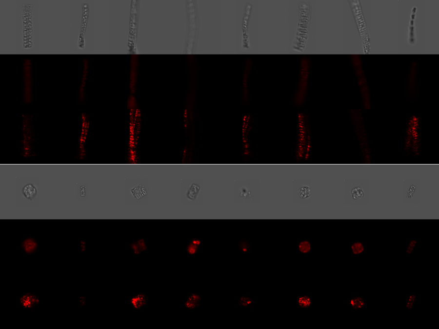
Phytoplankton biodiversity is a key indicator of marine ecosystems health. Due to climate change derived impacts, phytoplankton communities worldwide are being affected by changes in the water temperatures and the access to nutrients and light. Current methods to quantitatively classify phytoplankton samples require taxonomy experts to manually count and classify the individual cells they see […]
BetaSeg
Volume electron microscopy is the method of choice for the in situ interrogation of cellular ultrastructure at the nanometer scale, and with the increase in large raw image datasets generated, improving computational strategies for image segmentation and spatial analysis is necessary. Here we describe a practical and annotation-efficient pipeline for organelle-specific segmentation, spatial analysis and […]
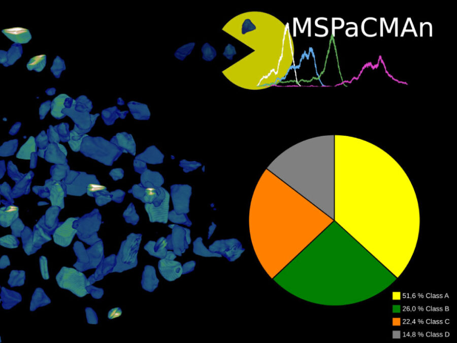
MSPaCMAn
MSPaCMAn is a workflow for quantifying mineral phases in 3D images of particulate materials using X-ray computed micro-tomography, designed to minimize imaging artifacts. It involves dispersing particles into samples, optimizing image processing to label individual particles, and identifying phases at the particle level by interpreting the grey-values of all voxels within each particle. This method […]
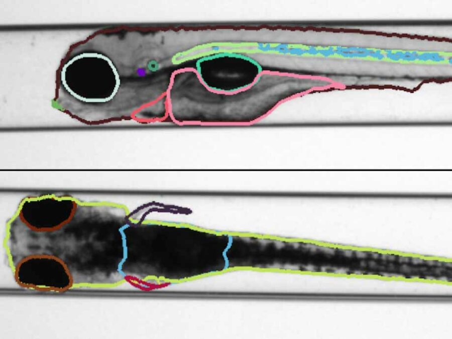
Identifying structural features of zebrafishes, using Semantic Segmentation
Zebrafish have a certain genetic similarity to humans and vertebrates. Particularly, the use of embryos is attractive due to the small scale screening capacity. Furthermore, similar to genuine cellular in vitro approaches, zebrafish embryos are considered as alternatives to animal testing. Therefore, they play a fundamental role in the detection of environmental and human health […]
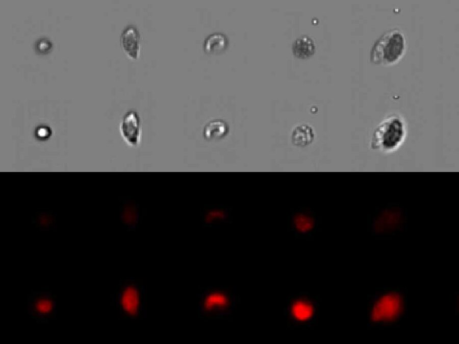
Automated Analysis of Evolutionary Experiments of Phytoplankton
There is a strong interest in understanding community assembly and dynamics. Experimental approaches using phytoplankton have proven to be extremely insightful to unravel underlying biological processes. Imaging flow cytometry is an emerging method becoming more and more popular in different fields. It allows us to capture changes within a community or population in a more […]
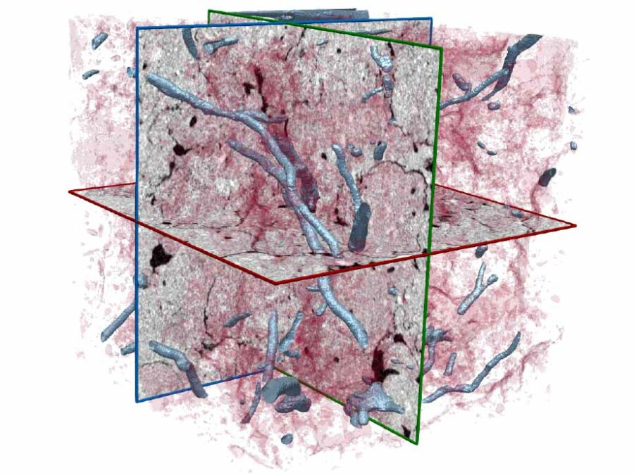
Understanding and analyzing biopores and plant roots in Soil Cores using Semantic Segmentation of CT Images
To understand how processes in ecosystems work and how they are connected the analysis of soil systems is essential. Since traditional computer vision methods for analysing soil cores reach their limits the next step is to integrate deep learning methods. Therefore a sufficient amount of labeled ground truth data is needed. Since labeling this large […]
Pick Yolo
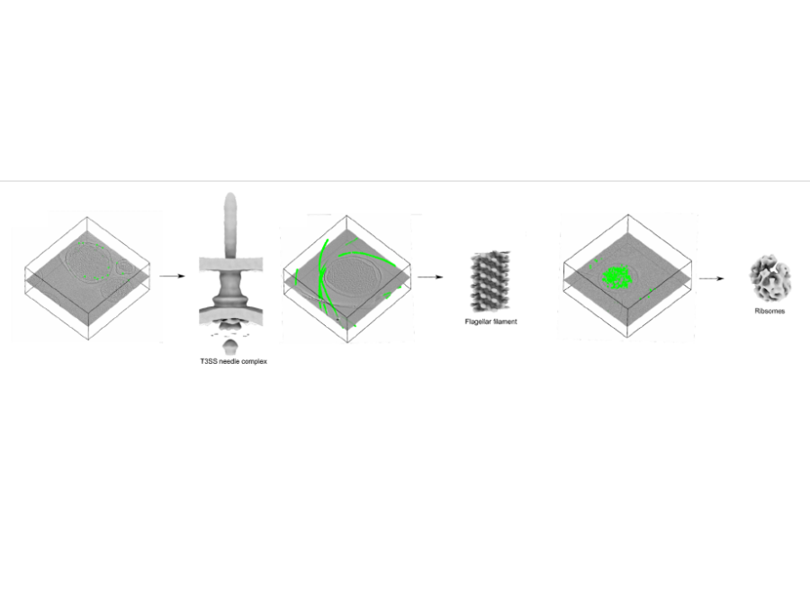
In collaboration with the Center for Structural Systems biology, Helmholtz Imaging has developed and trained a convolutional neural network for the picking of instances of proteins in cryoelectrontomograms (CryoET). The so picked instances are subsequently used to reconstruct a 3D structure of the proteins using subtomogram averaging methods. The novel picking methods exploit the well […]
Plaque Assays
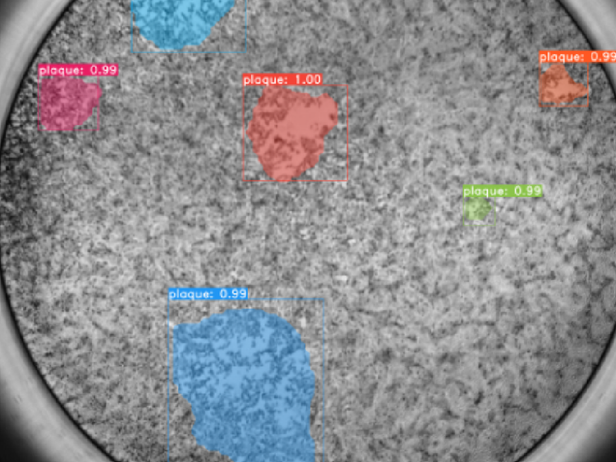
In collaboration with the Leibniz Institute of Virology and the Center for Structural Systems biology, Helmholtz Imaging is working on a processing pipeline for plaque assays. Plaque Assays are used to e.g. estimate the virus concentration in a given sample. For this high resolution images of cell cultures which have been infected with a virus […]
Segmentation of biodegradable bone implants
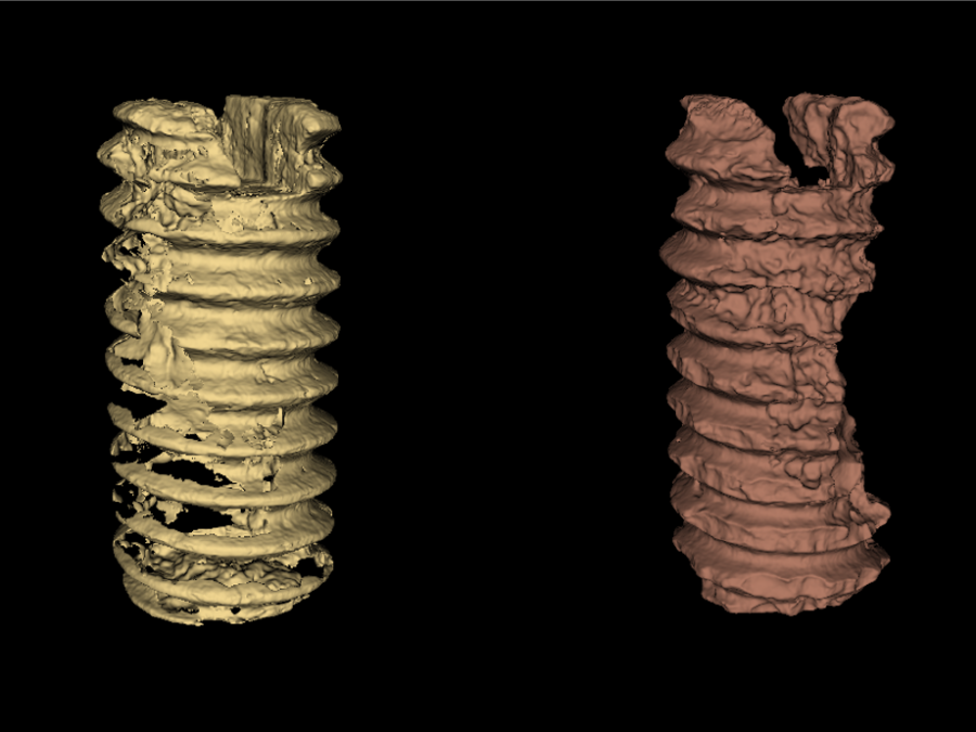
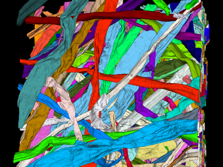
Together with the experts from Hereon the Helmholtz Imaging Support Team collaborates on segmenting synchrotron CT data. In 2021 the collaboration on semantic segmentation of biodegradable bone implants using a U-net resulted in two publications acknowledging the contribution of Helmholtz Imaging [1],[2]. Beyond that, we studied “Instance segmentation of paper fibers” imaged at the Hereon […]
Unisef
In the context of the Unisef project funded by Helmholtz AI, we implemented the prototype of a webservice to allow training and application of deep learning networks for segmentation running on the HPC infrastructure of DESY. The main idea here is to make DL based segmentation accessible and usable by non-experts, as well as complementing […]
Active Learning-enabled Generation of (Patho-)Physiological Lung Architectures for Pulmonary Medicine (ALEGRA)
ALEGRA aims to discover new insights into the morphology, physiology, and functionality of mammalian lungs. Therefore, quantitative parameters will be extracted from light sheet fluorescence microscopy images featuring comprehensive annotations of unprecedented detail for structures of interest such as hollow airways, blood vessels, as well as alveoli. These annotations are made possible by a novel […]
Connecting membrane pores and production parameters via machine learning (COMPUTING)
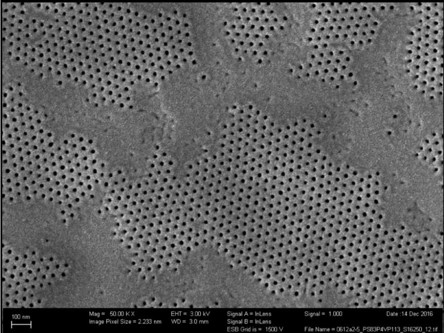
Isoporous block-copolymer membranes play a fundamental role in the filtration of liquids and can, for example, be used to purify drinking water. Despite recent progress in understanding the membrane formation process, finding suitable production parameters for a given precursor material (such as polymers of a certain length) still occurs in a trial-and-error fashion, wasting materials, […]
Detecting Type-2 Diabetes in histopathological images for a better understanding of biological processes behind the disease
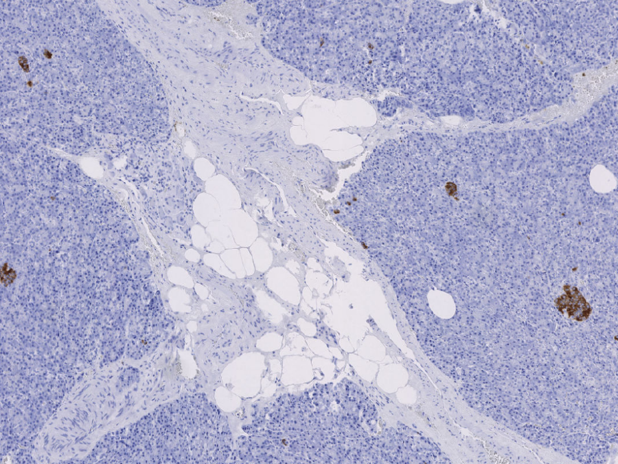
Type-2 diabetes is a chronic disease affecting about 500 million people worldwide. Despite extensive research over the last decades, exact biological processes leading to a deteriorating insulin production are not yet fully understood. By building models that are able to classify whether a patient has type-2 diabetes or not from whole slide images of the […]
