BRLEMM
Breaking resolution limit of electron microscopy for magnetic materials
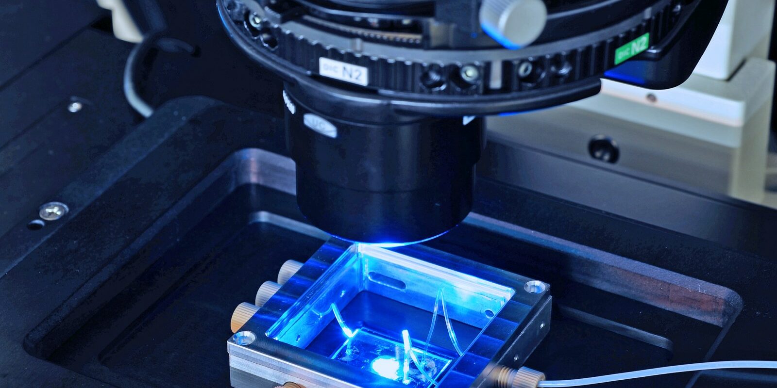
Iron-rhodium is the metal that Prof. Dr. Christian Kübel and his colleagues are mainly working with at present. Placing it under an electron microscope, Christian Kübel of the Karlsruhe Institute of Technology explains, “At room temperatures, it’s antiferromagnetic. When you increase the temperature, it becomes ferromagnetic.” The transition between these two states is a research area of great interest: how do defects in the atomic structure or stresses around precipitates influence the magnetic structure and thereby affect the magnetic properties of the metal?
The trouble is that it has been difficult so far to precisely quantify how much myelin is present. Experienced radiologists can assess brain tissue subjectively from MRI scans, where the variations in contrast allow them to draw conclusions about the state of the myelin, however they cannot determine the exact amount.

This is precisely the challenge to be tackled in this project. “We want to use a ptychographic approach to directly reconstruct the phase gradients, rather than the phase,” Christian Kübel says. This involves scanning samples under an electron microscope to deliver so-called diffraction pattern data. Instead of taking the usual approach of reconstructing phase and amplitude information from this data to calculate the magnetic properties, which is subject to error, the researchers with the group of Frank Filbir of the Helmholtz Zentrum München want to develop an algorithm that computes the magnetic information directly from the data. This is an important step forward for the basic research of magnetic materials, given that the method will be useful not only for iron-rhodium but also for many other materials.
Other projects
AsoftXm
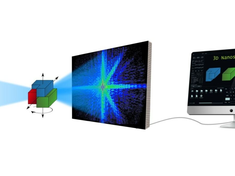
Advanced Soft-X-Ray Microscopy Solutions
The project aims to develop a method that will speed up the analysis of diffraction patterns that arise in UV and soft X-ray light microscopy, so that the structure of the studied sample can be calculated more efficiently. The method could make the three-dimensional study of nanomaterials considerably easier. There are times when researchers need […]
SATOMI
Tackling the segmentation and tracking challenges of growing colonies and microbialdiversity
An artificial intelligence will observe the growth of bacteria: from microscope images of bacterial cultures taken at regular intervals, it will precisely track the development and division of individual cells – even when multiple bacterial species are cultivated together.
SyNaToSe
Leveraging Cross-Domain Synergies for Efficient Machine Learning of Nanoscale Tomogram Segmentation
The aim is to develop an adaptable algorithm that can be used to perform different tasks in data and image analysis without needing to be trained with new, laboriously annotated images for each separate task.