MULTI-TRACE
Simultaneous quantitative multi-molecular PET imaging of two radiopharmaceuticals: from algorithmic developments to applications in neuroscience and oncology
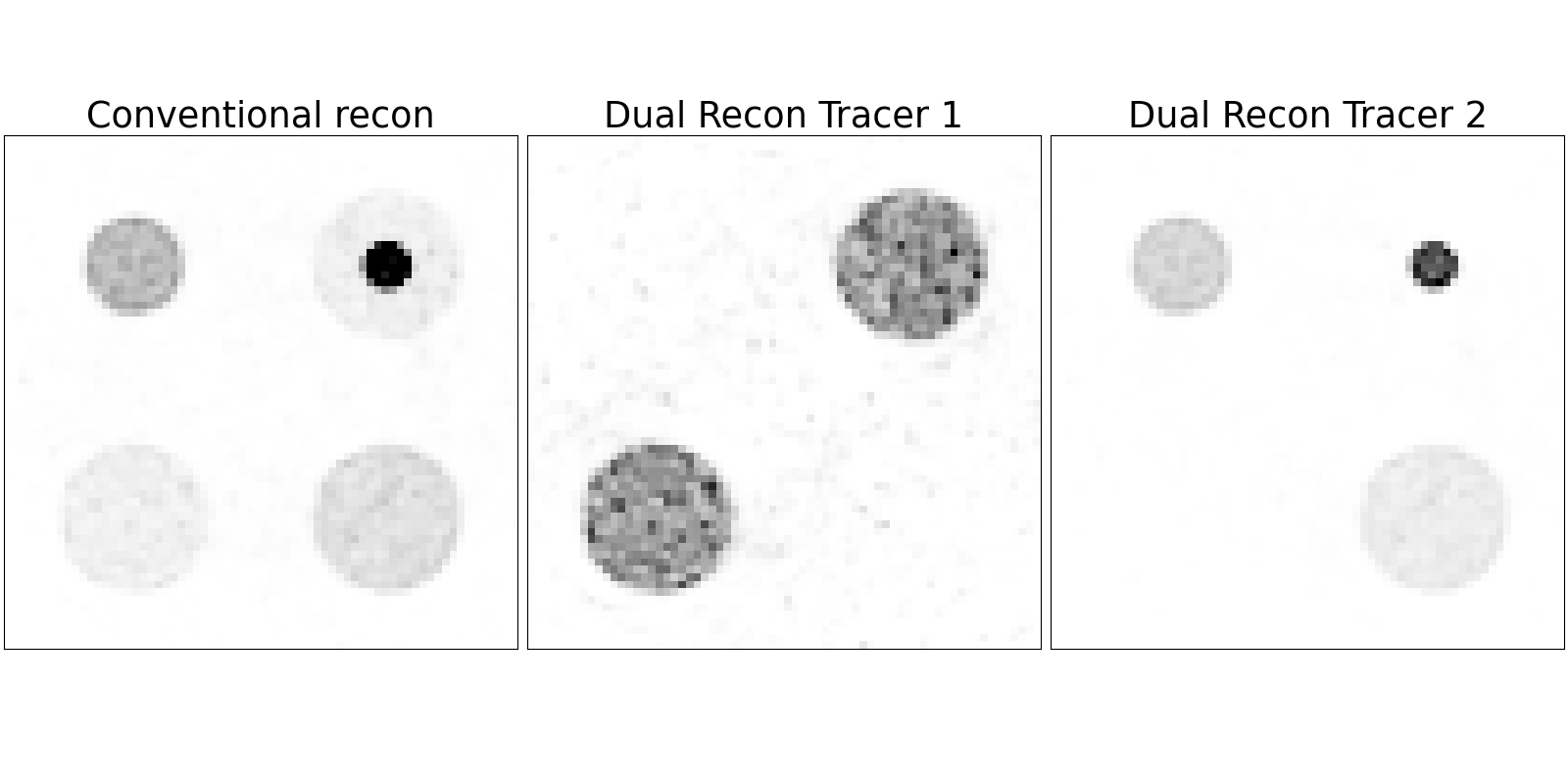
Positron emission tomography (PET) is a widely used imaging technique in neuroscience, oncology and pharmaceutical research, enabling the quantification of molecular processes using specific radiotracers. Currently, PET investigations are typically limited to a single tracer per scan, restricting insights to one molecular process at a time.
MULTI-TRACE aims to overcome this limitation by developing novel algorithms that enable the simultaneous detection and quantification of two different radiotracers within a single PET scan. The software will be compatible with both research and clinical PET systems and a range of radiopharmaceuticals, including tracers whose production will be established within the project. Released as a user-friendly open-source tool, the approach is expected to significantly advance PET imaging in research and clinical practice.
Other projects
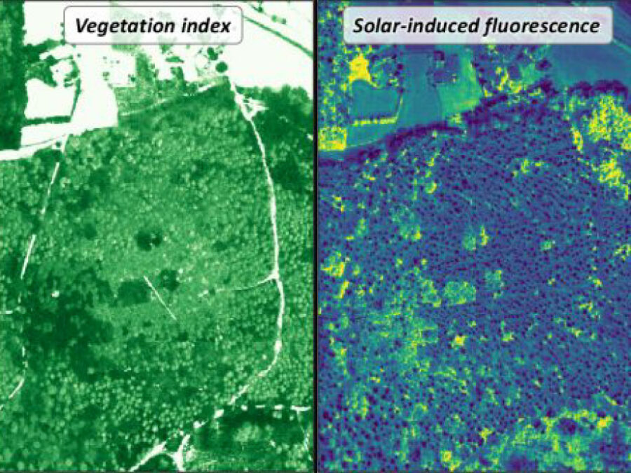
3DforestSIF
Understanding the solar-induced fluorescence (SIF) signal of natural, complex tree canopies
3DforestSIF seeks to correct airborne solar-induced fluorescence (SIF) data from forests for canopy structural and illumination effects, providing valuable insights for the early detection of forest stress.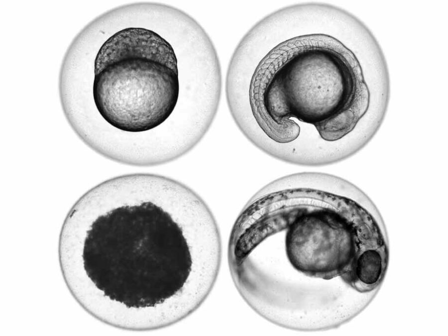
ImageTox
Automated image-based Detection of Early Toxicity Events in Zebrafish Larvae
ImageTox wants to establish an automated image-based system to assess zebrafish larval development. This will allow for a fast and unbiased evaluation of pathophysiological events during toxicological studies. To achieve this, the imaging process has to be optimized and a reliable model for sequence recognition based on deep learning has to be developed.
UTILE
Autonomous image analysis to accelerate energy materials discovery and integration
Research into green materials for clean energy generation is moving at full speed – yet still requires a long time to complete. This project is working on an open source image processing application that uses artificial intelligence to drive the analysis and management of image data from experiments across the energy materials community.