Katharina Kriegel
Published on 30.07.2026
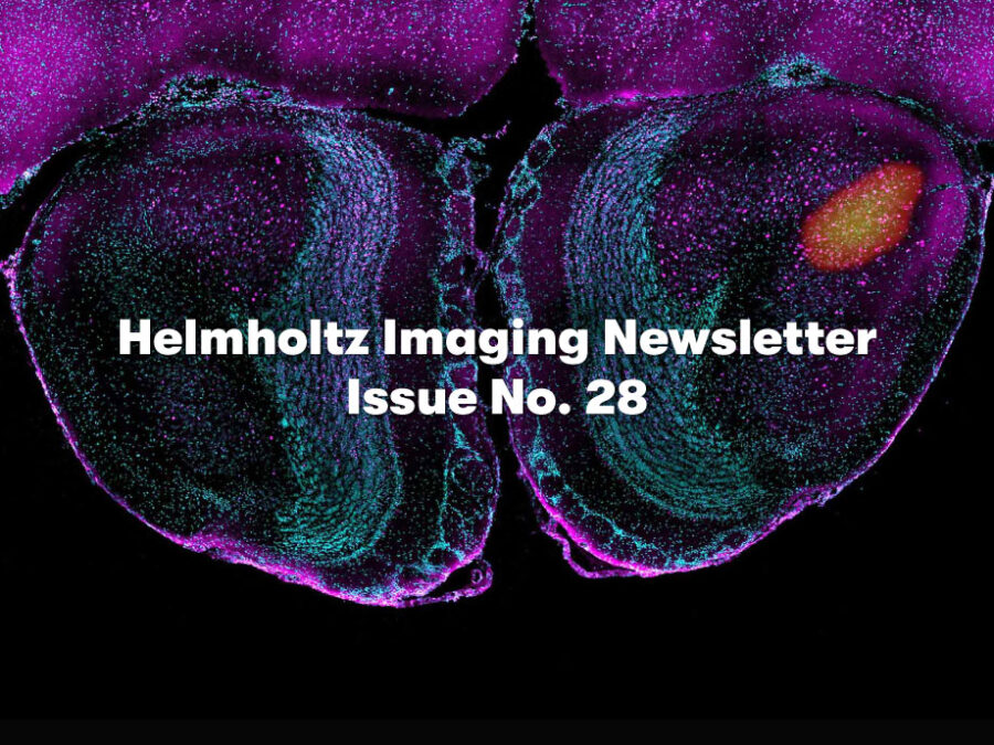
Helmholtz Imaging Newsletter Issue No. 28
Explore this year’s Best Scientific Image Contest entries, register for Helmholtz Imaging Week 2026, discover our new projects, and tools, and catch up on training opportunities and community updates in our latest newsletter.
Published on 27.07.2026
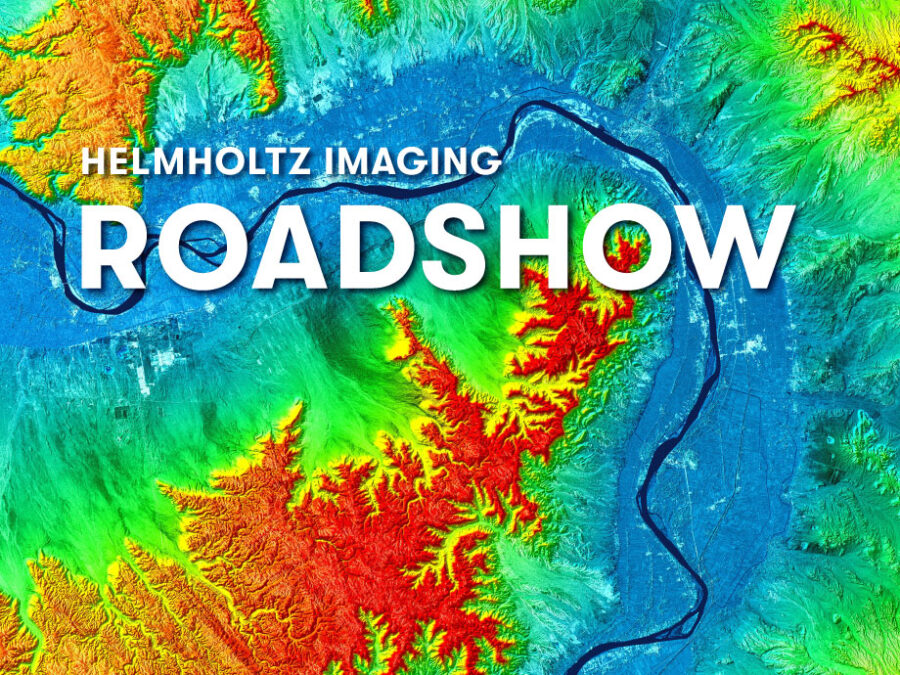
Bring your Imaging Questions: Helmholtz Imaging Roadshow at DLR
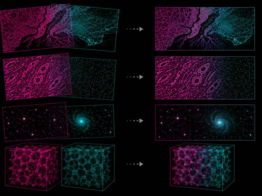
Join us on September 1 at DLR in Cologne for the Helmholtz Imaging Roadshow! Bring your imaging-related questions and discover how we can support your research. While you’re there, explore the winning images from our 2025 Best Scientific Image Contest, which will be on display on campus.
Published on 27.07.2026
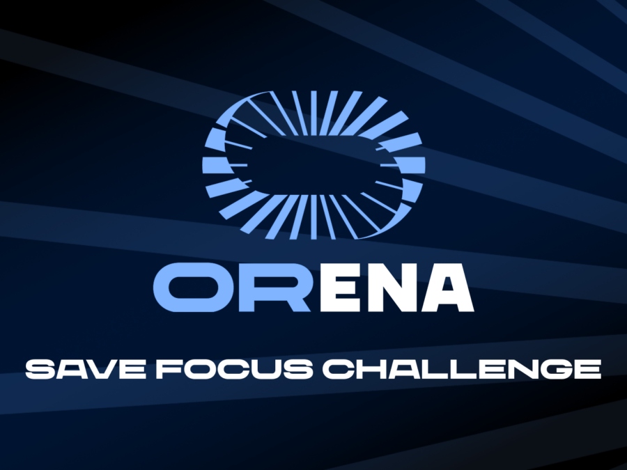
ORena SAVE FOCUS Challenge tests VLM’s surgical video understanding
Can vision-language models help surgeons keep track of every surgical object during an operation? The ORena SAVE FOCUS challenge puts that question to the test. Teams are invited to train their models on expert-annotated surgical video data and compete against the baseline models and each other. A $60k prize pool will be distributed among the top performers!
Published on 23.07.2026
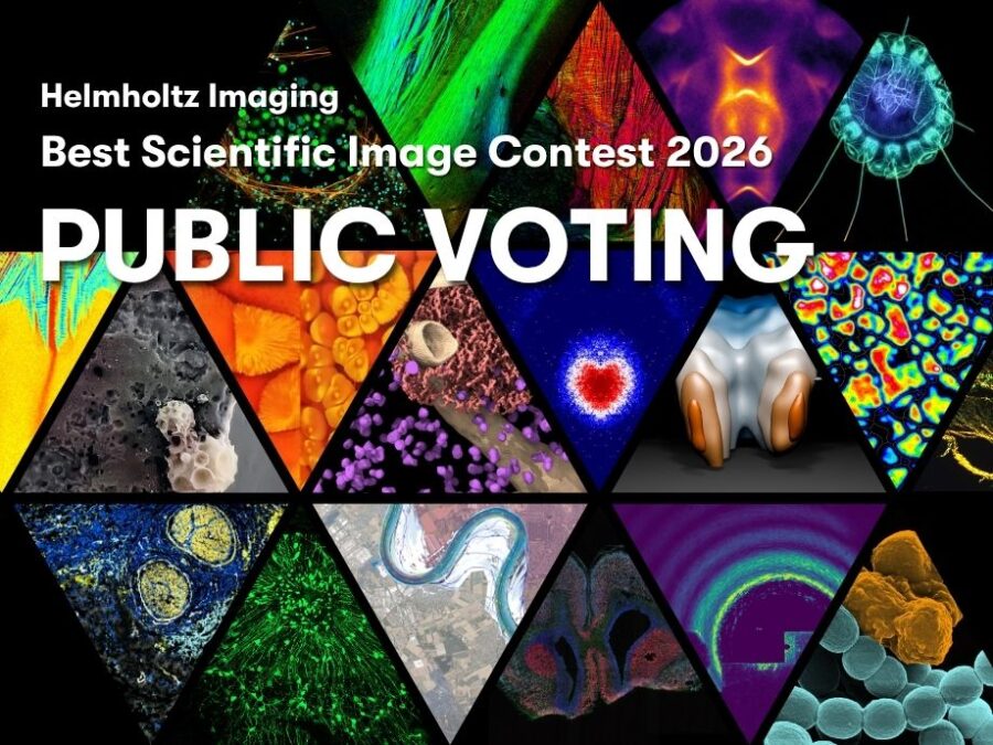
Cast Your Vote for the Public Choice Award
The images are in – and now it is your turn to choose your favorite scientific images for the Public Choice Award in the Helmholtz Imaging Best Scientific Image Contest 2026. Voting will open on July 30. Stay tuned!
Published on 14.07.2026
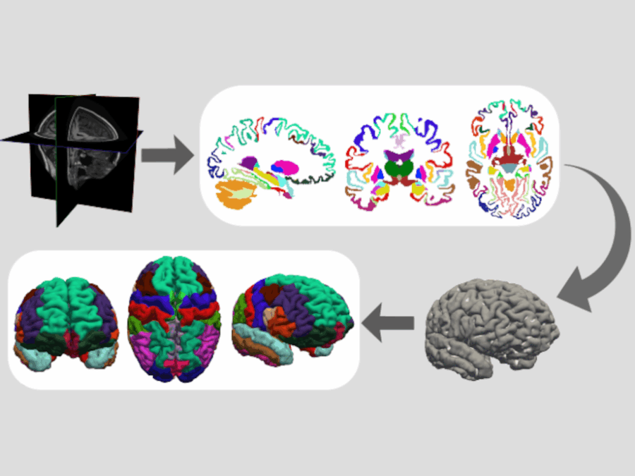
DeepMI FastSurfer / FreeSurfer course 2026
Join this 2.5-day, hands-on, introductory course on state-of-the-art deep-learning methods for fast and reliable neuroimage analysis from October 14 to 16 in Bonn!
Published on 26.06.2026
Helmholtz Imaging moves to Matrix
We are moving our community chat from Mattermost to Matrix. The new Helmholtz-wide platform, hosted by HIFIS, will support secure exchange and collaboration across the Helmholtz Association. Join us!
Published on 23.06.2026
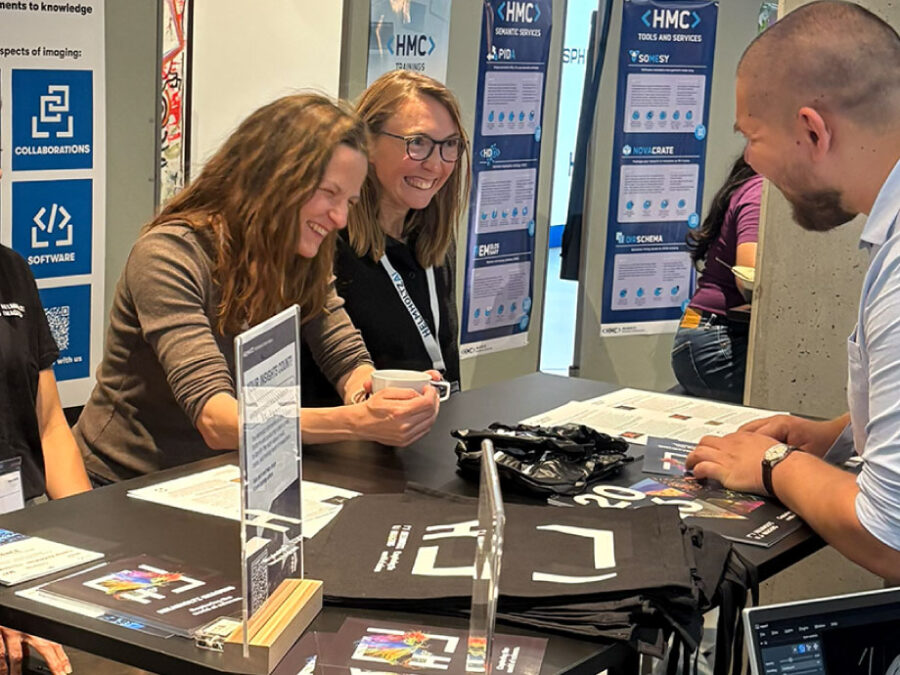
Helmholtz Imaging at HAICON26: Connecting Imaging, AI and Scientific Discovery
With hands-on workshops, a busy booth, and a poster spotlight on the Helmholtz Model Zoo, Helmholtz Imaging had many opportunities at the conference to connect with AI-savvy researchers and discuss how we can lift their imaging-related research to the next level.
Published on 18.06.2026
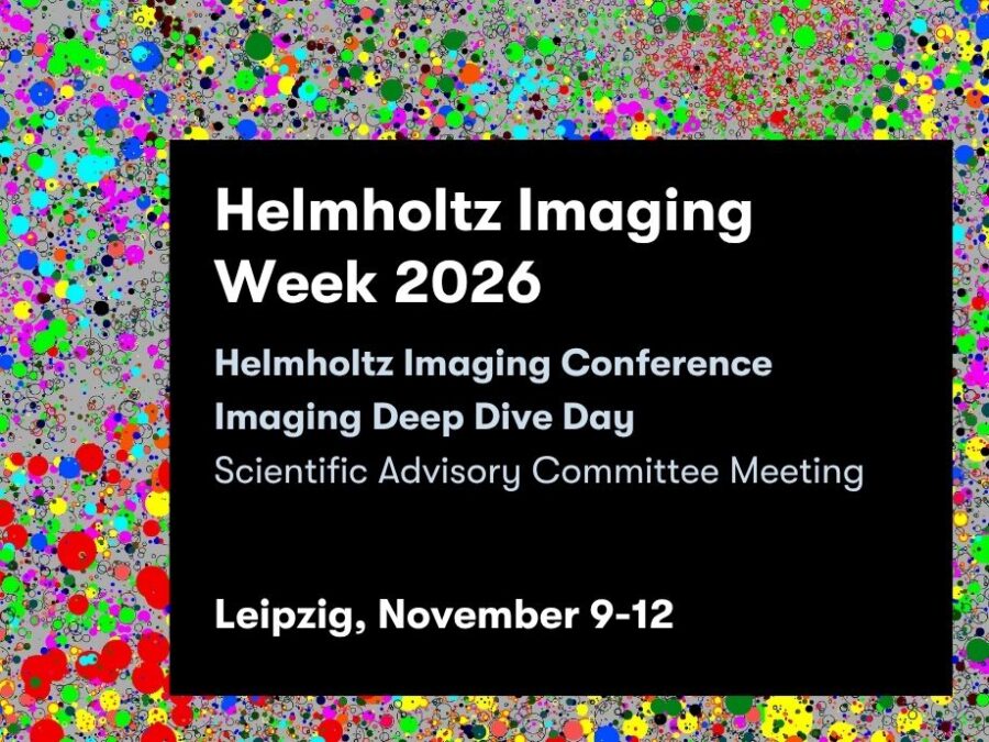
Helmholtz Imaging Week 2026
Mark your calendars: Helmholtz Imaging Week 2026 is coming to Leipzig from November 9–12, 2026, bringing together the imaging community for an extended program of scientific exchange, collaboration, and inspiration. Join us for the Imaging Deep Dive Day & annual Helmholtz Imaging Conference! Registration is open now.
Published on 18.06.2026
Introduction to Image Registration
Want to compare image data, align datasets, or apply integrated image registration techniques? This 90-minute workshop on November 4 introduces the essentials and practical techniques for image registration. Registration opens on October 7,

Published on 17.06.2026
Image Dataset Quality Control, Data Exploration and Pixel Patrol
Explore practical ways to assess image dataset quality, visualize metadata, and prepare data for downstream research with PixelPatrol. Join us virtually on September 10 & 17! Registration opens August 13.
Published on 16.06.2026
Helmholtz Imaging is moving to Matrix
Helmholtz Imaging is moving its community communication from Mattermost to Matrix. Join our new Matrix space for community updates, exchange, and sharing. Mattermost will be shut down by July 1, 2026.

Published on 16.06.2026
Open Call: ScienceServe, Boosting Research Software at Helmholtz
Apply for project funding to enhance the sustainability and quality of research software! Deadline: August 14, 2026

Published on 04.06.2026
Survey: Help Shape Helmholtz’ Data Science Training Portfolio
Participate in a short survey on current training needs and future skill development priorities!
Published on 29.05.2026
Helmholtz Imaging Newsletter Issue No. 27
From the Project Call 2026 and upcoming training opportunities to new community tools and a preview of Helmholtz Imaging Week 2026, our latest newsletter highlights what’s happening across Helmholtz Imaging and beyond.
Published on 19.05.2026
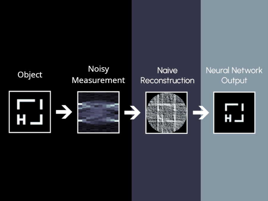
Regularization in Image Reconstruction: From Model to Data Driven Methods
Build a strong foundation in image reconstruction and inverse problems in this 7-hour online course on September 16, 2026. Registration opens August 19.