Helmholtz Imaging Projects
Published on 26.06.2026
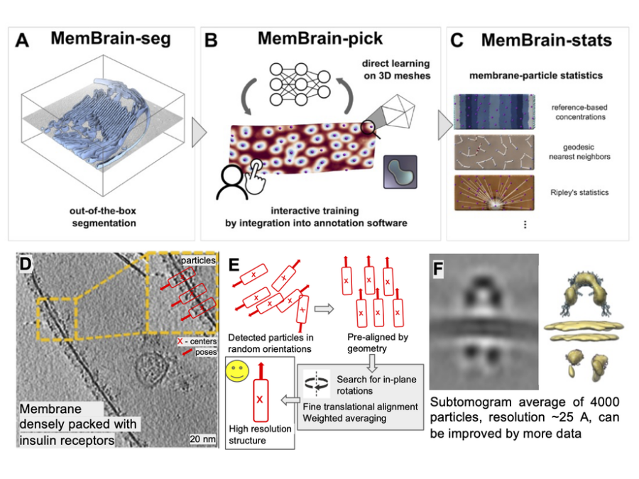
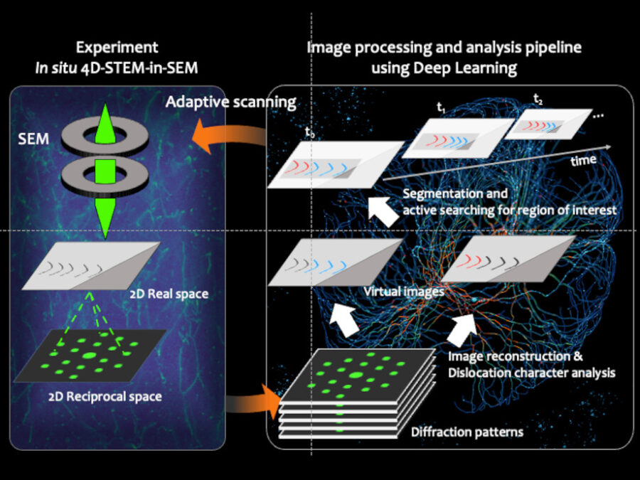
MemBrain-structure
An open-source AI pipeline automates cryo-ET data analysis, turning raw tilt-series into 3D membrane protein structures relevant for biology and medicine.
Published on 18.06.2026
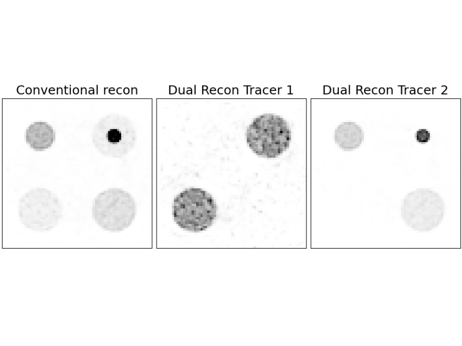
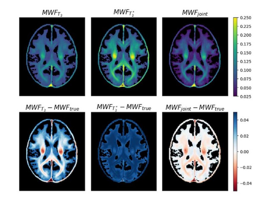
MULTI-TRACE
New open-source algorithms enable dual-tracer PET imaging, allowing two molecular processes to be studied simultaneously in a single scan.
Published on 15.06.2026
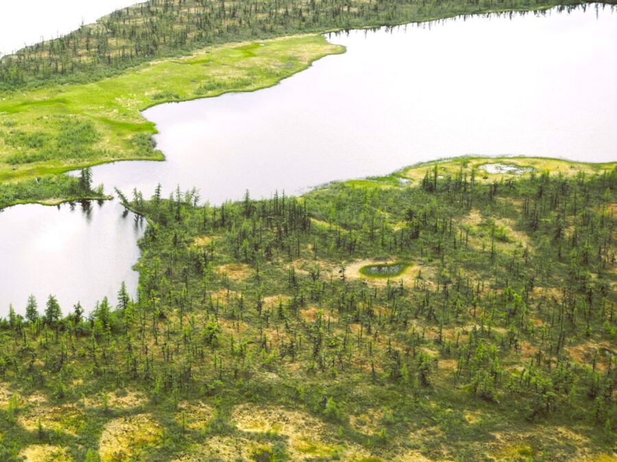
Moisture3D
Drone-based radar imaging and AI reconstruct 3D soil moisture underground, enabling improved monitoring of water resources and environmental processes.

Published on 27.05.2025
PlastoView
Water quality is essential for ecosystems and human health, yet it’s increasingly threatened by microplastics. This project develops image-based methods for detecting both plankton and microplastics using a new low-cost, mobile system.
Published on 19.05.2025
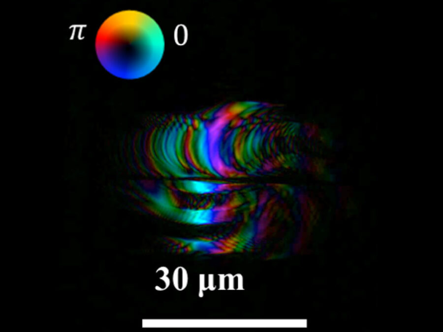
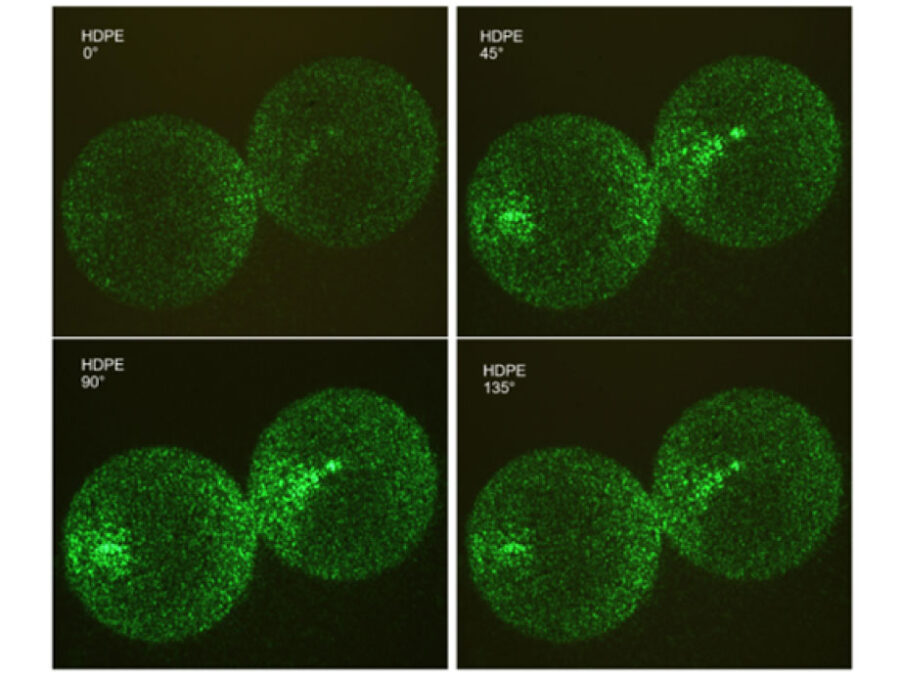
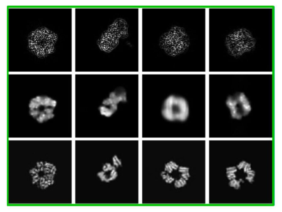
cryoFocal
This project explores how defocused images recorded with an electron microscope can be used to reconstruct the 3D structure of molecules inside cells. This method aims to enable faster and more cost-effective structural analysis of molecules to accelerate understanding of their functions and to design drugs against them.
Published on 07.10.2024
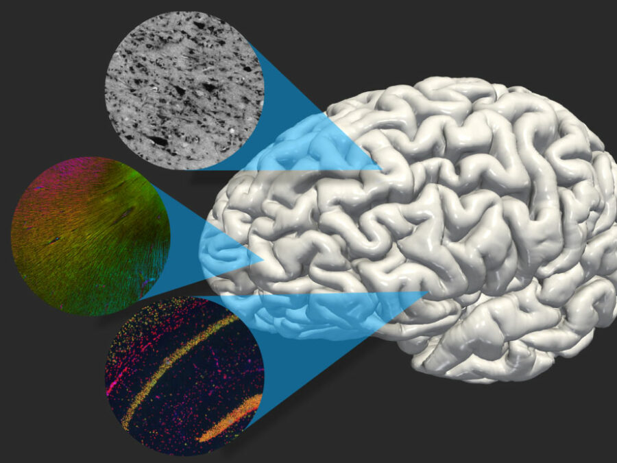
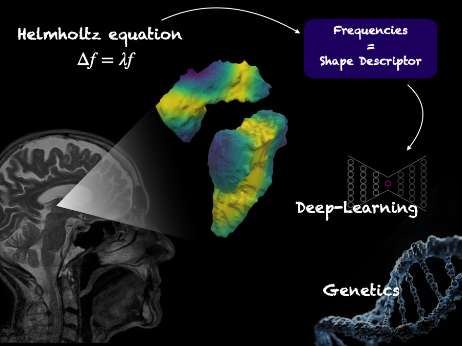
BrainShapes
The project explores the 3D structure of the human brain by creating a digital ‘map’ of the brain and examining its unique genetic properties, potentially linking genetic variations to brain disorders.
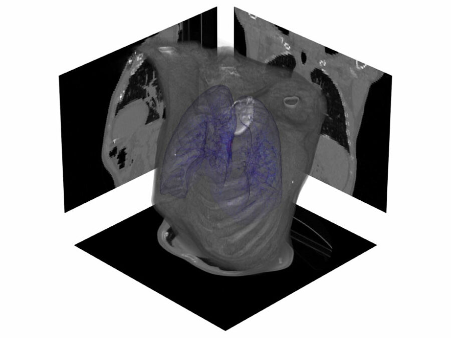
Published on 19.08.2024
CLARITY
Dose-escalated radiotherapy of lung cancers requires precise monitoring of lesions and nearby organs at risk. Current methods are able to track ultra-central lesions but neglect their deforming vicinity, risking unacceptable toxicity to aortico-pulmonary structures. AI-based anomaly detection and generative AI models can address both requirements in real-time.
Published on 14.08.2024
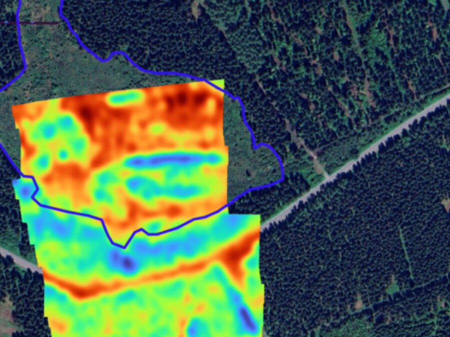
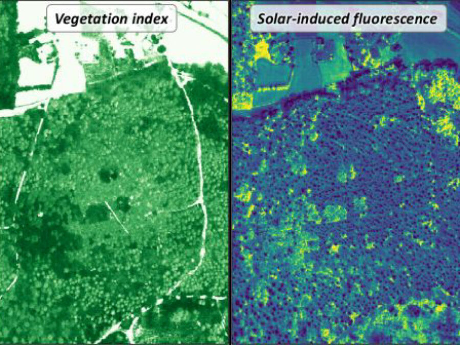
3DforestSIF
3DforestSIF seeks to correct airborne solar-induced fluorescence (SIF) data from forests for canopy structural and illumination effects, providing valuable insights for the early detection of forest stress.