Best Scientific Image Contest 2026
The sixth edition of the Best Scientific Image Contest showcased again the fascinating diversity of scientific imaging across the Helmholtz Association. With 125 impressive submissions, there was plenty to discover! Explore the top 21 images in our virtual gallery and uncover the science behind them.
Gallery

Insights into the Developing Heart (1st place Jury Award)
Alena Boos & Nadine Spielmann, Helmholtz Munich
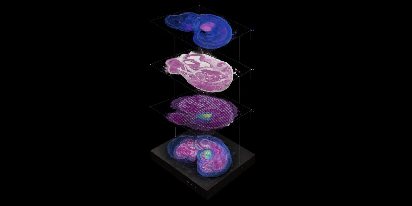
This image unfolds the developing mouse heart across four connected layers: a fluorescent view of the whole embryo, a stained tissue section, a color-coded map of gene activity, and a final reconstruction that brings all information together. By aligning anatomy, tissue structure, and gene expression in the same spatial framework, researchers can see where specific genes are active as the heart forms. Comparing healthy and genetically modified embryos may reveal how disruptions in these carefully coordinated processes lead to congenital heart defects, supporting future advances in diagnosis and treatment. Captured using whole-mount fluorescence microscopy, brightfield microscopy of H&E-stained tissue, and spatial transcriptomics.
Enterprise: Keyhole Encounter Insights (2nd place Jury Award & 1st Place Participants' Choice Award)
Paul H. Kamm, Helmholtz-Zentrum Berlin, Fraunhofer Institute for Material and Beam Technology IWS & TOMCAT beamline at the Swiss Light Source
Resembling a starship inside molten metal, this image shows a laser-welding keyhole, a narrow, vapor-filled channel, glowing red as it pierces a pressurized gas cavity. Blue bubbles emerge within the gray weld seam, capturing a highly dynamic process that can create pores and weaken welded components. This is the first three-dimensional observation of such a keyhole in action, revealing how molten metal, gas, pressure, and surface tension interact within milliseconds. Understanding these processes helps researchers optimize laser movement, reduce defects, and produce stronger, safer welds in die-cast aluminum. Captured using operando synchrotron X-ray tomoscopy, producing a complete 3D image every 10 milliseconds at a voxel size of 2.75 µm. Access publication: https://doi.org/10.1002/advs.202413108
Carbonate Rainbow (3rd place Jury Award)
"Saeid Asadzadeh, GFZ Helmholtz Centre for Geosciences "
This colorful satellite view reveals carbonate rock layers in the Anti-Atlas Mountains, with blue tones indicating dolomite-rich areas and red tones marking calcite-rich rocks. Intermediate colors show gradual changes in composition across strata that preserve hundreds of millions of years of Earth history. By mapping subtle chemical variations from space, researchers can investigate ancient fluids, temperatures, and geological processes that transformed these rocks over time. Information once available mainly through laboratory analysis can now be traced continuously across entire landscapes, turning invisible chemistry into a geological story in color. Captured from orbit using EnMAP satellite-based hyperspectral optical remote sensing, which records reflected sunlight in more than 200 spectral bands.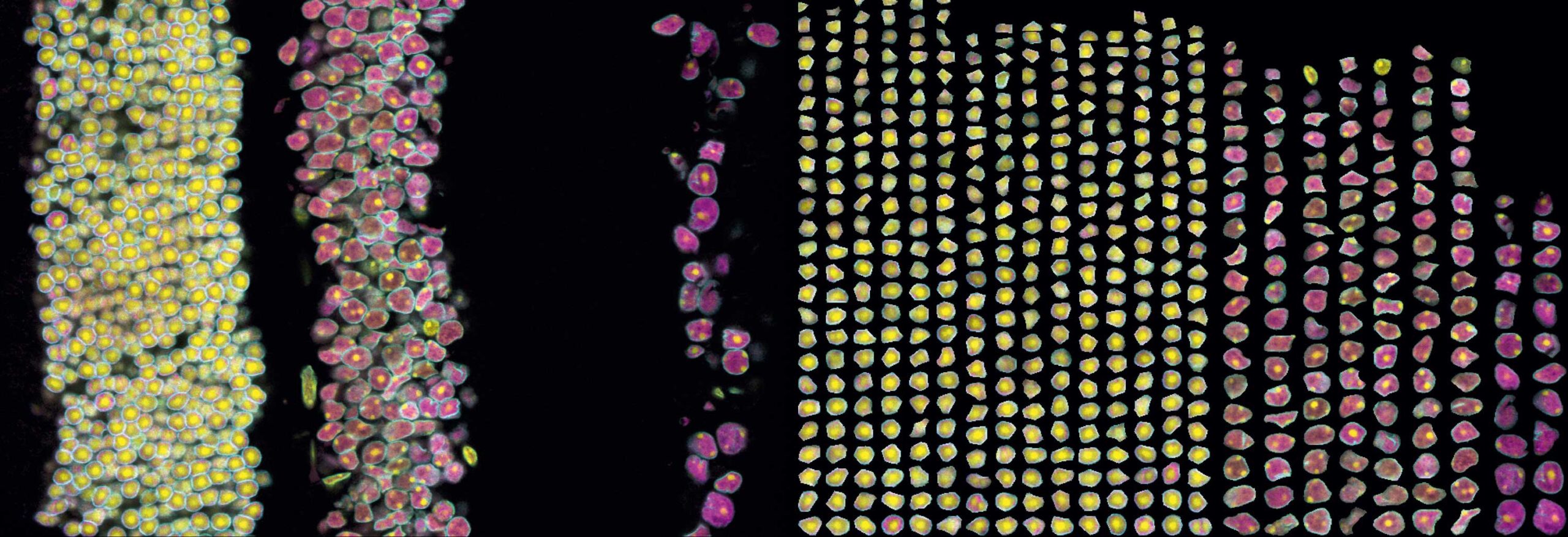
The Art of Nuclear Order (1st place Public Choice Award)
Meghna Joon & Johanna Kroll, MDC
This image shows a cross-section of mouse retina with distinct cell layers on the left and individual nuclei from each layer neatly arranged on the right. The colors reveal the nuclear lamina in cyan, chromatin in yellow, and newly synthesized RNA in magenta. By computationally separating and reorganizing the nuclei, differences between retinal cell types become immediately visible. Of particular interest are rod photoreceptors, whose nuclei in nocturnal animals have an unusual, inverted chromatin organization. Comparing these different nuclear architectures helps researchers understand how the spatial arrangement of chromatin influences transcription and gene activity across cell types, with relevance for development, adaptation, and disease. Captured using confocal fluorescence microscopy with a Plan Apo 63×/1.4 NA oil objective.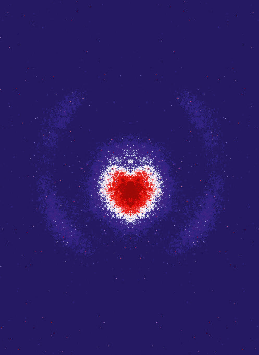
The Beauty of Broken Symmetry (2nd place Public Choice Award)
Valerija Music & Markus Ilchen, DESY, University of Hamburg
Thousands of individual electron hits unexpectedly form a heart-shaped pattern in this raw experimental image. In an ideal velocity-map measurement, electrons with identical velocities would create perfectly symmetric circular distributions. Here, tiny changes in electric fields and alignment break that symmetry, turning microscopic deviations into a visible structure about 1.5 centimeters wide. The pattern appeared during the first successful test of a newly built spectrometer at a synchrotron facility, using a photon beam to ionize a gaseous target, and was not created by image processing. Beyond its striking appearance, the asymmetry provides useful diagnostic information for improving measurement precision. The instrument is designed to study ultrafast electron dynamics in chiral molecules. Captured using double-sided electron velocity-map imaging with an MCP/phosphor detector and CCD camera readout.
Fireworks in the Eternal Darkness (3rd place Public Choice Award)
Veit Dausmann, Anton Theileis, Tobias Strickmann & Jan Taucher, GEOMAR
A large jellyfish drifts through the darkness 2,500 meters below the surface of the Mozambique Channel. Artificial-intelligence-based coloring indicates its position and extent within the sampled water volume, transforming a rare deep-sea encounter into measurable data. Due to their delicate morphology, jellyfish are difficult to survey in the vast deep ocean and may make up a larger share of zooplankton than previously thought. Images like this help researchers map their distribution, measure body size, estimate biovolume, and improve calculations of carbon movement through the ocean. The same imaging approach supports large-scale assessments of plankton biodiversity and particles. Captured with the PIScO extended-depth-of-field shadowgraphy system, developed at GEOMAR for in-situ deep-sea plankton imaging.
Two Paths of Development (2nd Place Participants' Choice Award)
Nadine Spielmann & Gerhard Przemeck, Helmholtz Munich
Two mouse embryos from the same litter reveal dramatically different developmental paths: one has developed normally, while the other is undergoing resorption after development was disrupted in a model of congenital heart disease. The contrast captures both the precision and fragility of embryonic development. Researchers use such images to document abnormalities, assess embryo viability, and test whether genes suspected from patient data can causally interfere with development. Comparing these outcomes helps clarify how disruption of essential genes can influence both heart formation and survival, strengthening the link between genetic changes and congenital disease. The embryos were freshly dissected and left unstained. Captured using brightfield stereomicroscopy.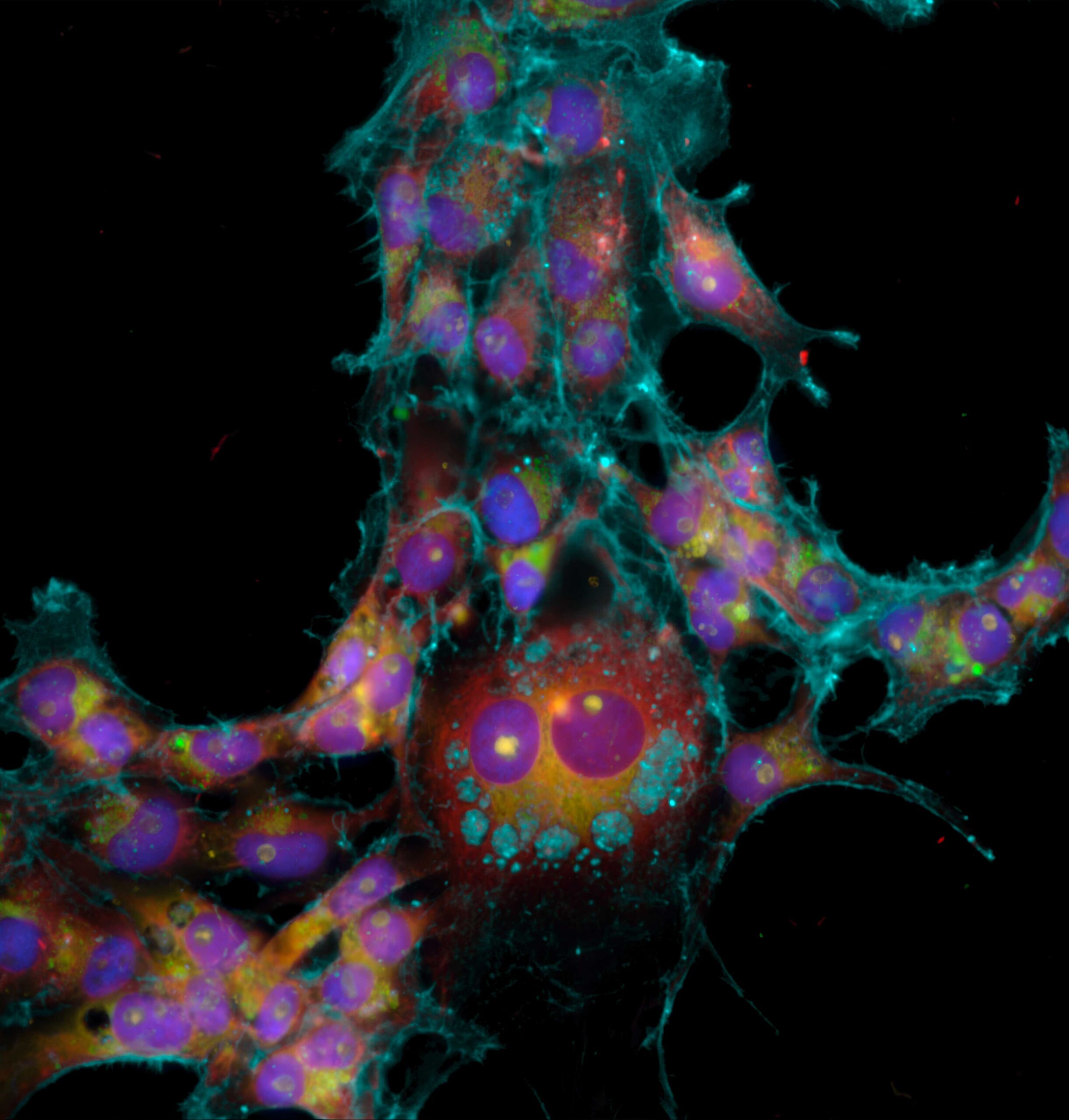
When Your Cells Smile Back at You (3rd Place Participants' Choice Award)
Luisa Reger & Jo Nyffeler, UFZ
Six fluorescent stains reveal the inner architecture of amphibian cells, with nuclei, RNA, organelles, and the delicate cytoskeleton forming a pattern that appears almost to smile back at the viewer. The beauty is deceptive: the cells were exposed to a chemical that alters their structure, including the actin cytoskeleton. This Cell Painting approach records many cellular features at once, allowing researchers to detect early signs of toxicity and compare how chemicals affect amphibian and other vertebrate cells. The method could improve amphibian-specific chemical risk assessment while reducing the need for animal testing. Captured using automated high-content fluorescence microscopy on an ImageXpress Micro Confocal system in widefield mode with multiple fluorescence channels.
Fermi Pig in an Iron Monolayer
Ying-Jiun Chen, FZ Jülich
This momentum-space image maps electronic states in an artificial crystal made from a single atomic layer of iron on a tungsten surface. Its most striking feature is a newly formed state nicknamed the “Fermi pig”, which exists in neither bulk iron nor bulk tungsten. It appears at the Fermi energy, where electronic states strongly influence properties such as electrical transport and magnetism. The feature reveals strong interaction, or hybridization, between iron and tungsten atoms at their interface and helps explain the material's unusual spin and charge transport. Such states could open new ways to use electron spin and orbital motion in future information technologies. Captured using momentum microscopy at Forschungszentrum Jülich.
Jellyfish-like Epithelial Cell
Mathias Müsken, HZI
Looking almost like a jellyfish in motion, this image shows a single epithelial cell isolated from mouse lung tissue. Removed from its usual tissue environment, the cell's body and delicate protrusions become visible in striking detail. Researchers use such images to investigate whether the shape and size of a specific group of lung epithelial cells change with age. Advanced image analysis and machine-learning methods can measure cellular dimensions and protrusions across different age groups, helping to reveal age-related changes in lung tissue at the level of individual cells. The cell was first isolated by fluorescence-activated cell sorting, then fixed and prepared for imaging. Captured using field-emission scanning electron microscopy with a Zeiss Merlin and colored in GIMP.
Microglial Engulfment
Cassandra Caedo, MDC
A yellow microglial cell with an amoeba-like shape sits within a dense network of human neurons. Cyan neuronal material is visible inside the cell, showing that it is engulfing and clearing parts of the surrounding network. Microglia are the brain's resident immune cells and continuously monitor their environment. This human stem-cell-derived culture brings separately developed neurons and microglia together, allowing researchers to observe their interactions directly. Images like this help reveal how microglia respond to neuronal material, remodel neural connections, and integrate into developing networks. Understanding these processes may shed light on neurodevelopment, neuroinflammation, and neuropsychiatric disease. Captured using confocal fluorescence microscopy.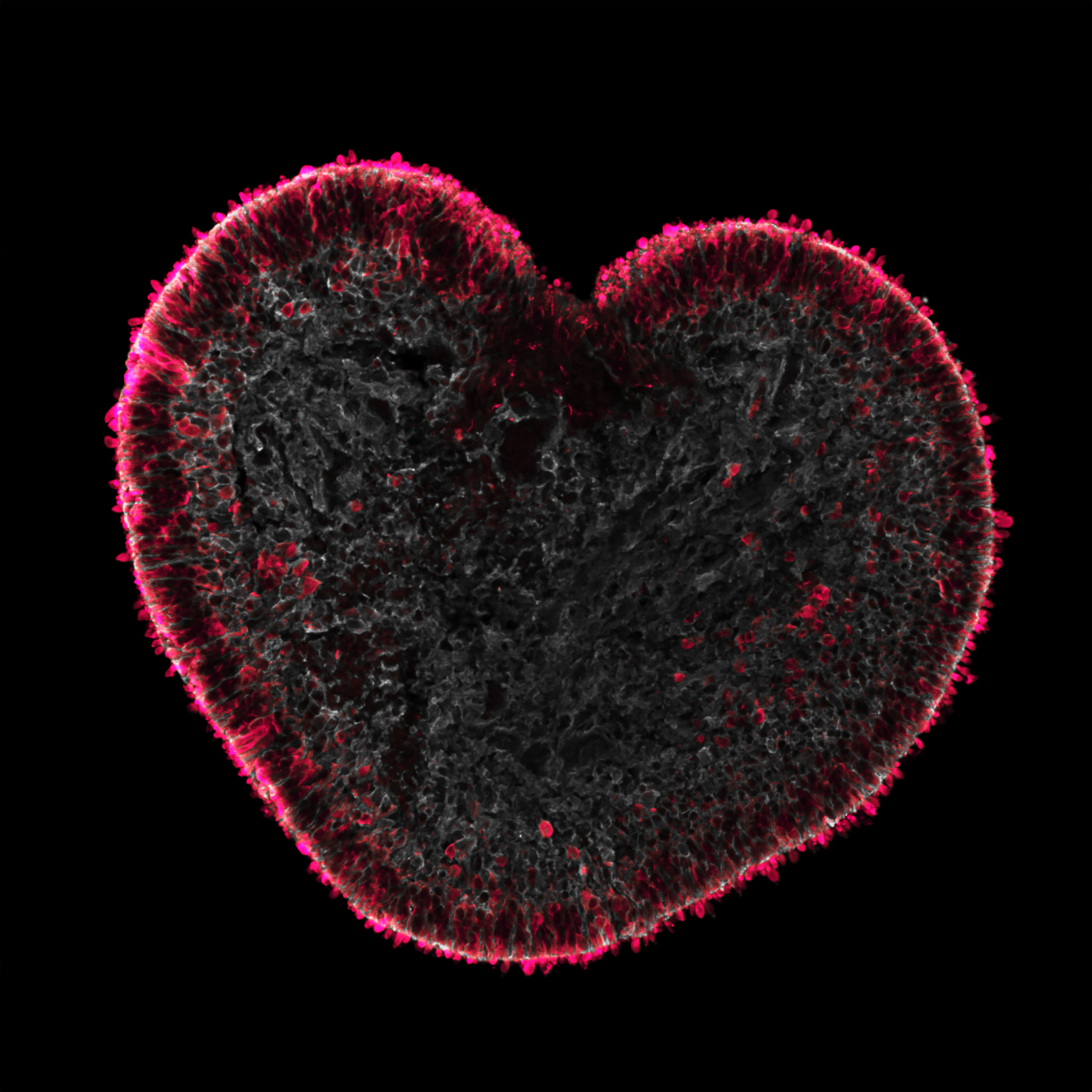
Love is in the Organoids
Natalie Dumler, DZNE
This unique heart-shaped structure is a human retinal organoid: a miniature, laboratory-grown model of the retina. Photoreceptor cells are labeled in magenta and Müller glia in white, recapitulating the retinal structure in vivo. The organoid comes from a line lacking PIEZO1, a calcium channel that senses mechanical forces, and formed part of an experiment exploring how biomechanics influence retinal tissue. Retinal organoids allow researchers to study processes involved in diseases such as retinal detachment and age-related macular degeneration without working directly in the human eye. They can also support the search for future therapeutic approaches. Captured using fluorescence microscopy on a Zeiss ApoTome 2 with 10x magnification and a maximum-intensity z-projection.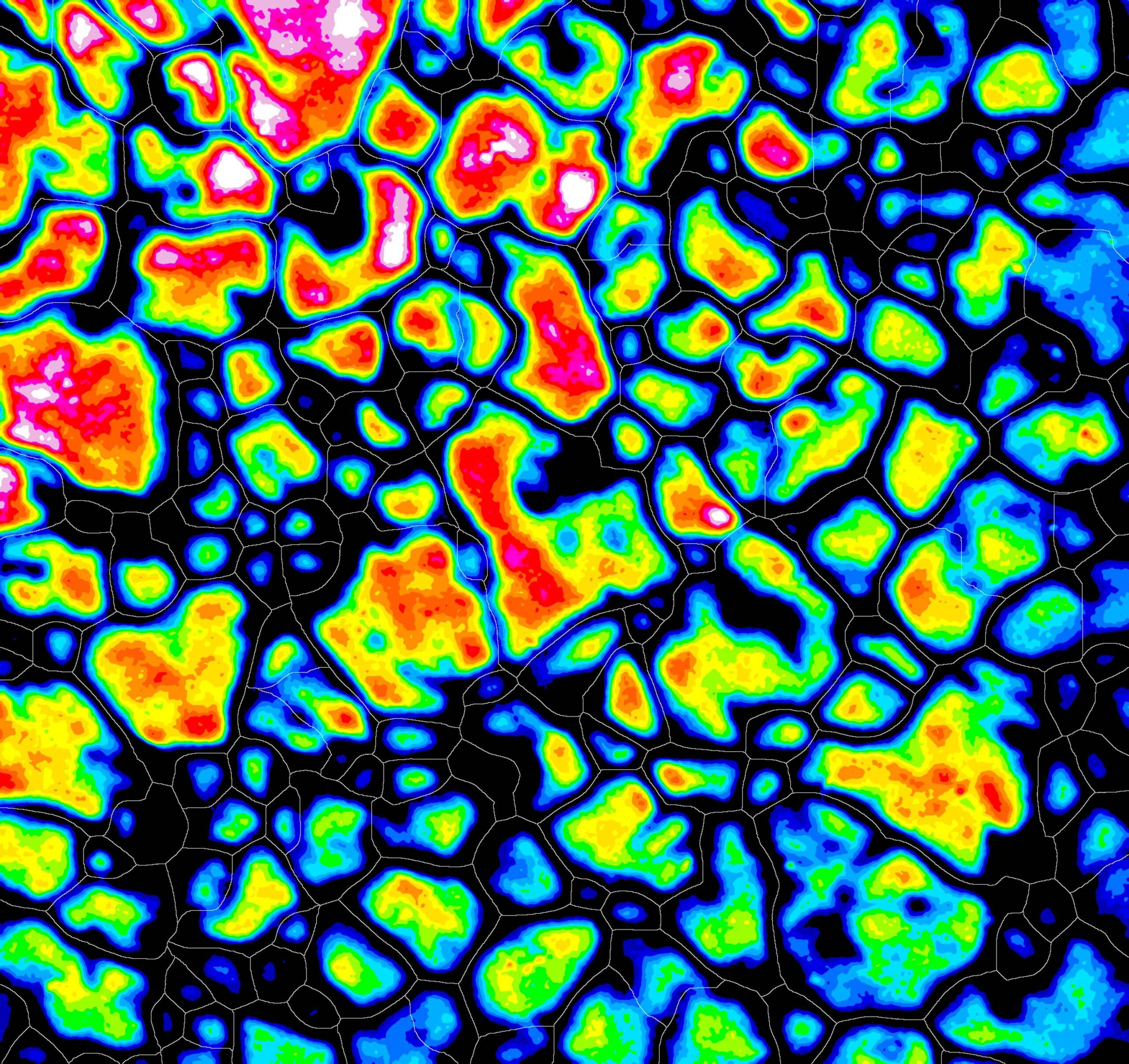
Fractured Crystals of Solar Energy
Yannic Kerkhoff & Daniel Abou-Ras, HZB
This color-coded image shows light emitted by a polycrystalline CuInSe2 absorber inside a finished solar cell. Bright grain interiors indicate stronger luminescence, while darker boundaries between crystals reveal areas where performance is reduced; thin white lines mark grains identified by a neural network. A nearly heart-shaped grain stands out near the center. By measuring luminescence at the scale of individual crystals, researchers can assess local material quality and determine how grain boundaries affect solar-cell efficiency. These insights help connect microscopic structure with device performance and guide the development of better photovoltaic materials. Captured by cathodoluminescence in a scanning electron microscope equipped with a mirror, spectrometer, and photon detector.
Tundra Snow Dragon
Inge Grünberg, AWI
A dragon seems to hide in this Arctic tundra valley: its legs and armored back are formed by areas of deep snow, shown in red, while violet and blue mark little or no snow. The pattern is more than a striking coincidence. Deep snow insulates the ground, warming the soil and reducing the stability of underlying permafrost. By mapping snow depth together with terrain and vegetation, researchers can identify areas especially vulnerable to thaw and improve models of how Arctic soils may respond to climate change. This matters because accelerating permafrost thaw can release additional carbon into the atmosphere. The map combines summer terrain and winter snow observations from Trail Valley Creek in Canada. Captured using airborne LiDAR on board an AWI polar aircraft.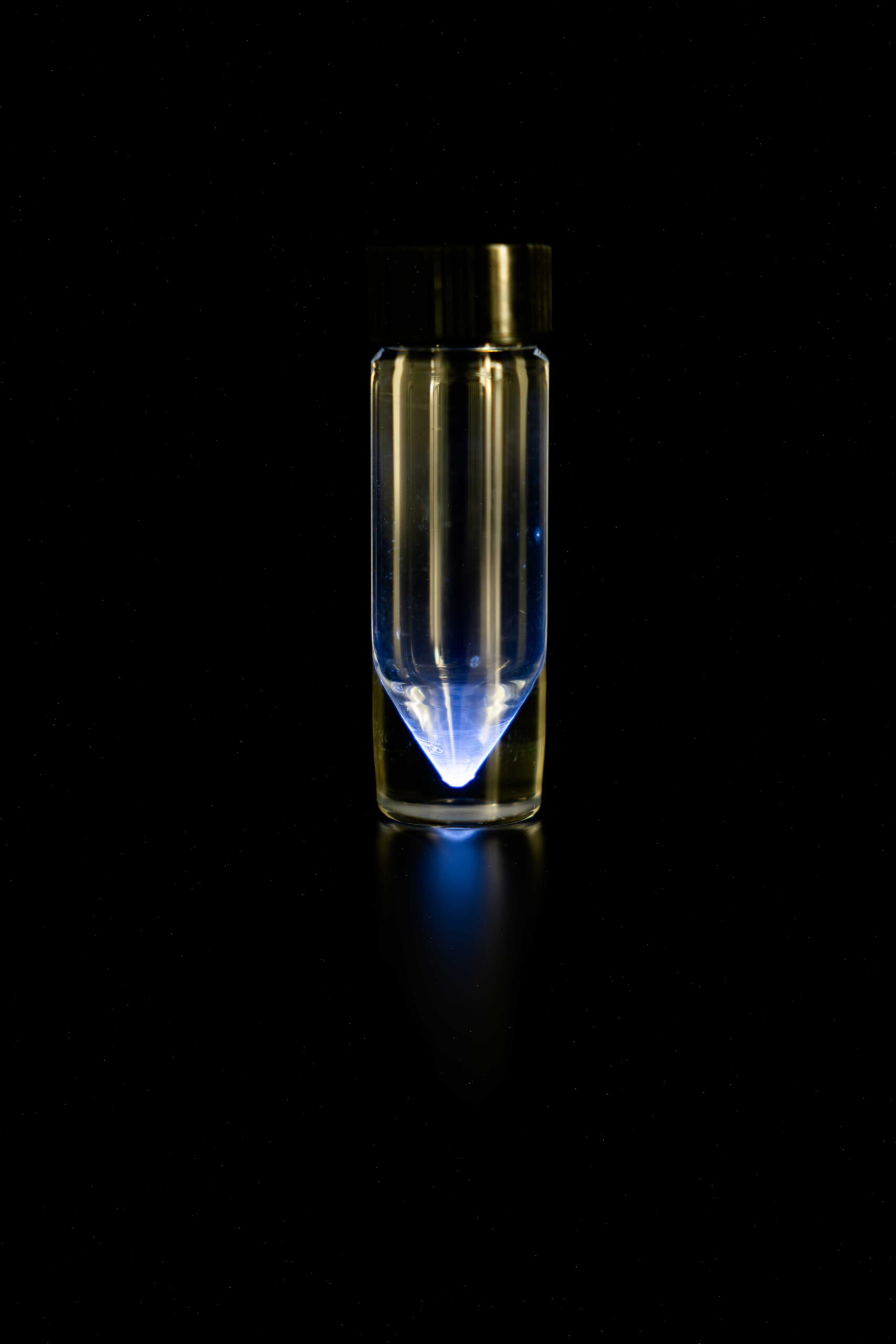
First Image of Astatine
Alexander Haseloer, FZ Jülich
A faint blue glow emerges from a tiny dried sample of astatine-211 inside a glass vial, the first indirect photograph of astatine described as the rarest naturally occurring element on Earth. The sample contained only about 40 nanograms, yet its intense radioactivity was visible in near-total darkness. The glow is thought to arise mainly when alpha particles from radioactive decay ionize the surrounding glass, rather than from added color or artificial lighting. Studying this radioluminescence provides clues to how radiation transfers energy to matter. Astatine-211 is also a promising radionuclide for targeted alpha therapy, where its short-lived, high-energy decay could be used to attack cancer cells. Photographed with a Canon R8 and RF24-70L F2.8 lens using a 106-second exposure.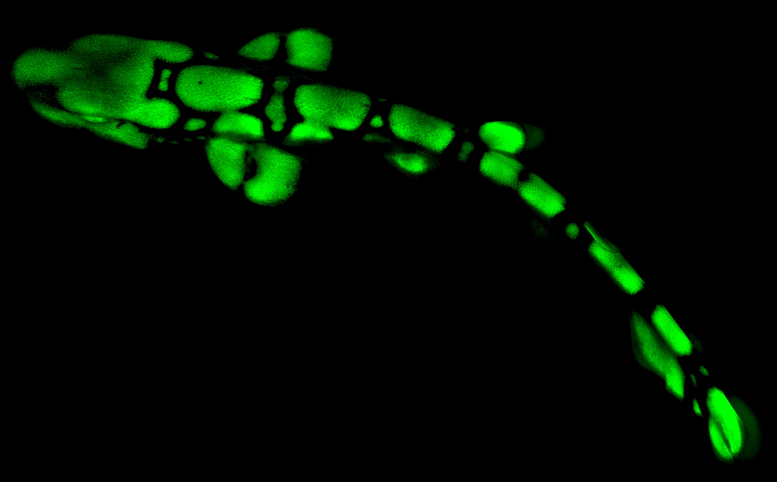
Sharkglow
Sampurna Chakrabarti, HZI
A chain catshark hatchling glows bright green under blue light, revealing a fluorescent pattern that would otherwise remain invisible. This biofluorescence is produced by small molecules derived from tryptophan and is rare even among sharks. Its purpose remains a mystery: one possibility is that the patterned glow helps chain catsharks communicate with one another while remaining less visible to other deep-sea animals. Studying this phenomenon can reveal how fluorescence evolved, what ecological role it plays, and which molecular mechanisms produce it. Such images make hidden traits of these ancient cartilaginous fish accessible to research, from biochemistry to behavior and evolution. Captured using an epifluorescence dissection microscope with a GFP filter; multiple images were stitched together.
Structural Plasticity of PspA Assemblies
Benedikt Junglas & Carsten Sachse, FZ Jülich
These rod-shaped assemblies of the bacterial protein PspA appear in a range of diameters, revealing a remarkable ability to change shape. PspA helps bacteria maintain and repair their cell membranes, but the underlying mechanism has long remained unclear. Structural data indicate that PspA can progressively constrict around membranes and shed small vesicles, with ATP providing additional energy for the diameter changes. Solving these structures was especially challenging because rods of different widths can occur within the same sample; researchers classified millions of particles and reconstructed more than 60 structures to build a consistent model. Captured using cryogenic electron microscopy on Titan Krios and Talos Arctica transmission electron microscopes with K3 direct electron detectors.
Neural Aurora
Marvin Edelmann, Franz Kärtner & Mikhail Pergament, DESY, Andreu Matamoros-Angles & Markus Glatzel, UKE
Cyan, magenta, and green fluorescence sweep across mouse brain tissue like an aurora, revealing several cellular and structural features at the same time. Each color represents a different fluorescent signal rather than a natural tissue color. Combining multiple labels in one image allows researchers to examine how distinct cell types, neuronal processes, and tissue structures are arranged in relation to one another. Two-photon microscopy is particularly useful for neuroscience because it can visualize fluorescent structures deeper inside biological tissue while reducing interference between spectral channels. Such images help build a more detailed picture of brain architecture and cellular organization in a three-dimensional environment. Captured using multicolor two-photon fluorescence microscopy.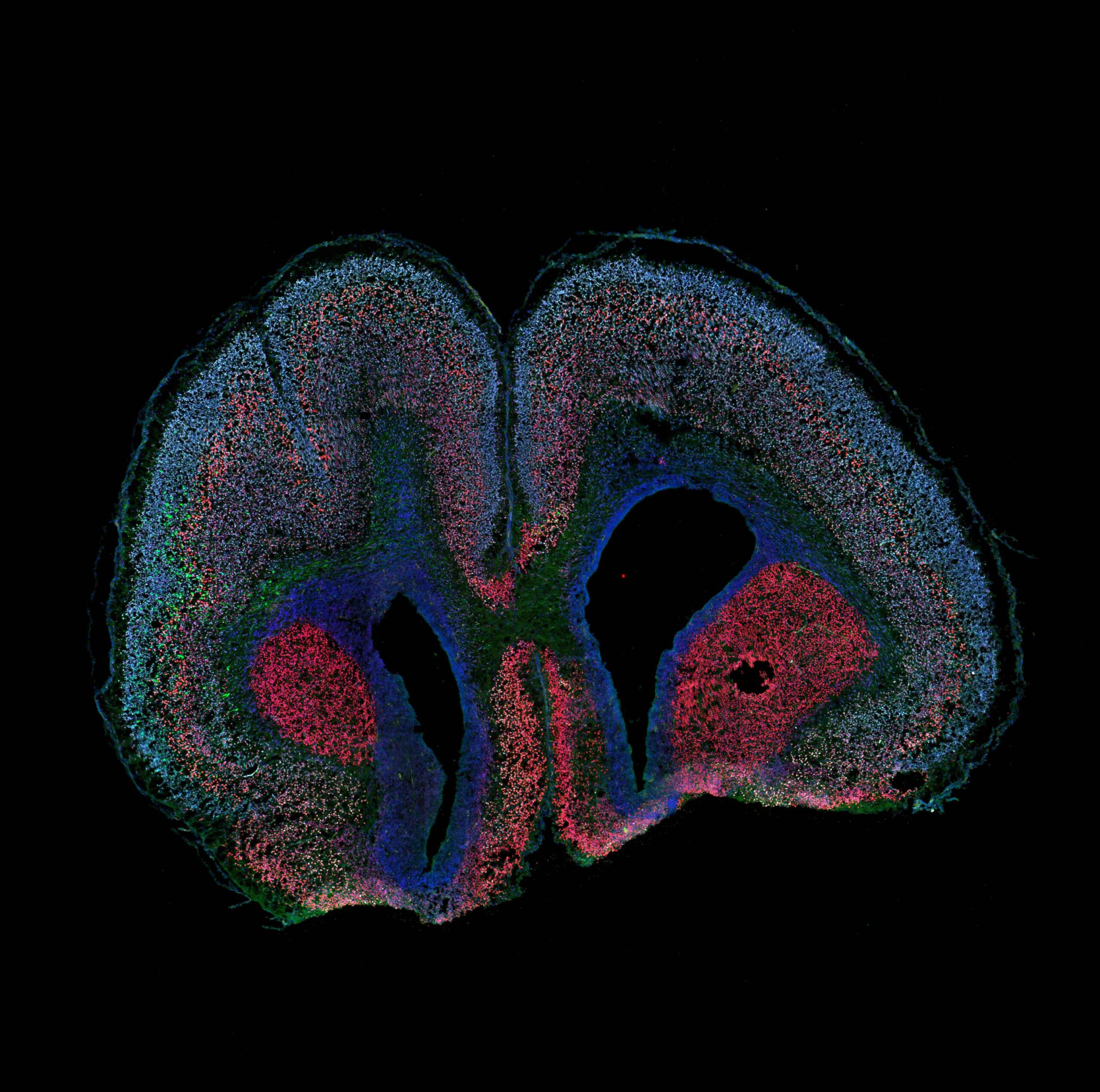
Neural Cartography
Anthi Krontira, Helmholtz Munich
This newborn mouse brain section resembles a cellular map: upper-layer cortical neurons appear gray, deep-layer neurons red, and genetically labeled cells green against the surrounding cortex and striatum. The ordered layers show the remarkable precision with which the developing brain is assembled. By introducing a genetic construct before birth and later tracking the position and identity of labeled cells, researchers can investigate how specific genes influence neuron formation, cell fate, and migration. These experiments help reveal how normal brain architecture emerges and how disruptions in developmental programs may contribute to neurodevelopmental disorders. Captured using spinning-disk confocal microscopy with a 40x water-immersion objective; multiple fields of view were stitched together.
Hot-as-hell
Bradley Guy, Jens Gutzmer, Tina Pereira & Samuel Thomas Thiele, HZDR
Cubic crystals of the mineral bixbyite cluster around a glowing cavity as if lit from within. The sample comes from South Africa's Kalahari Manganese Field, whose rocks preserve a remarkable record of Earth's history, including the rise of atmospheric oxygen and later the formation of the Supercontinent Rodinia. The study set out to identify tiny encrustations suspected to be the rare mineral saccoite. Instead, the coatings proved to be a complex mixture of carbonate and silicate minerals that formed after the bixbyite crystals. Even an unexpected result can therefore reveal how minerals formed and changed over geological time. Captured using an FEI Quanta 650F field-emission gun scanning electron microscope, combining backscattered and secondary electron signals with color gradients.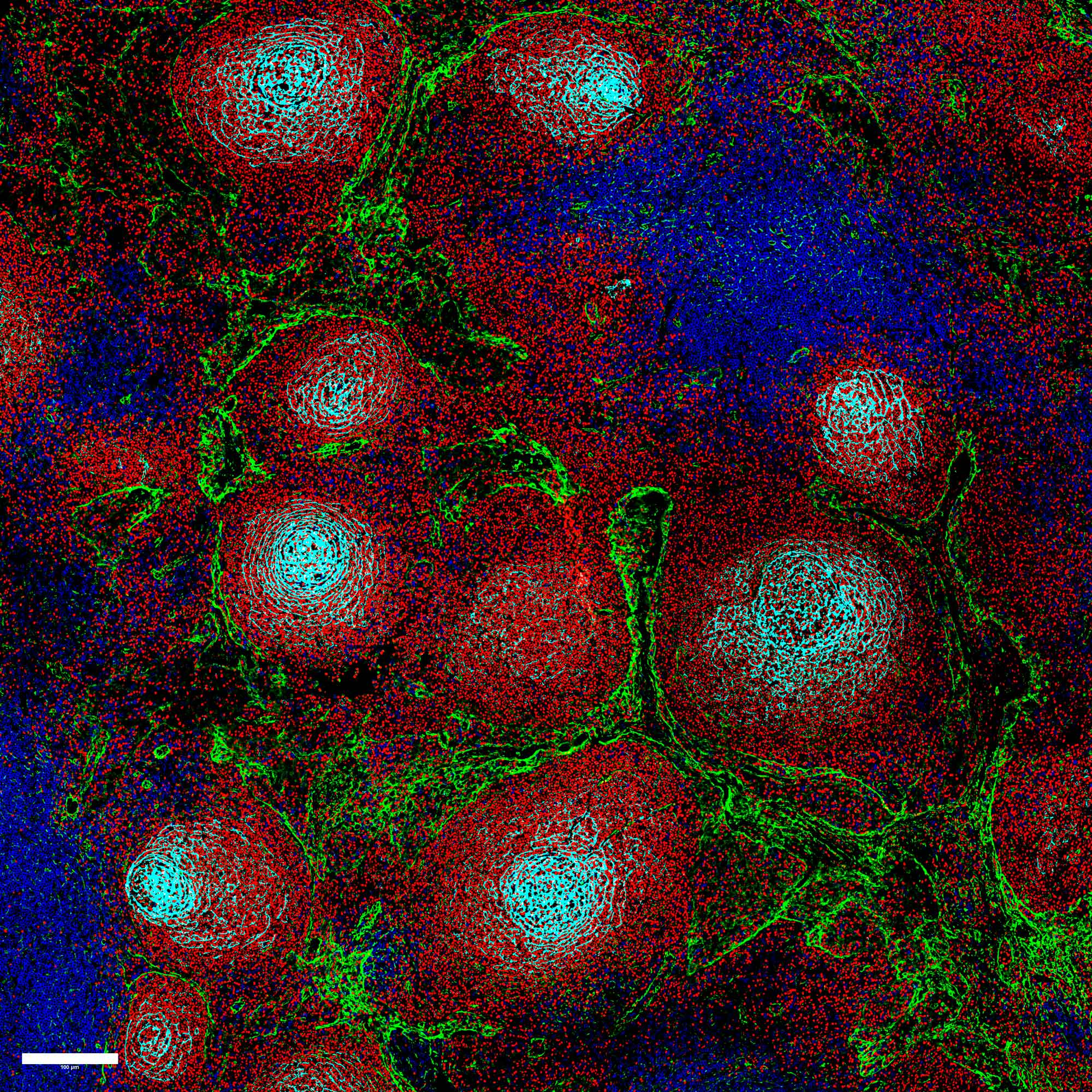
Human Lymph Node Immune Architecture
Daniel Hübschmann, Lea Jopp-Saile & Anna Mathioudaki, DKFZ, Felix Czernilofsky, Tobias Roider & Marc-Andrea Baertsch, University Hospital Heidelberg, Garry P. Nolan, Stanford University, USA, Judith B. Zaugg, University of Basel and University Hospital Basel, Basel, Switzerland, Simon Haas, MDC, Sascha Dietrich, University Hospital Düsseldorf
This multicolor image maps the architecture of a human lymph node at single-cell resolution. B cells appear red, T cells blue, and supporting stromal and follicular dendritic cells green and cyan, revealing clearly separated but closely connected cellular neighborhoods. This spatial organization is essential for coordinating normal immune responses. By comparing healthy lymph nodes with tissue transformed by lymphoma, researchers can investigate how these finely tuned structures become progressively disrupted during disease. The underlying experiment can detect 53 cellular markers, allowing immune and structural cell populations to be studied in remarkable detail and linked with other single-cell data. Captured using CODEX multiplex immunofluorescence imaging with a 53-antibody panel and a Keyence BZ-X710 fluorescence microscope. Access paper: https://doi.org/10.1038/s43018-026-01136-z