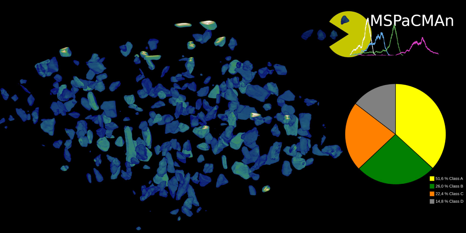
MSPaCMAn

Visual, HI Collaboration MSPaCMAn
Image: Dr. Jose Ricardo da Assuncao Godinho, HZDR, Jan Philipp Albrecht, MDC

MSPaCMAn is a workflow for quantifying mineral phases in 3D images of particulate materials using X-ray computed micro-tomography, designed to minimize imaging artifacts. It involves dispersing particles into samples, optimizing image processing to label individual particles, and identifying phases at the particle level by interpreting the grey-values of all voxels within each particle.

This method also incorporates particle geometry and microstructure for classification, improving phase classification accuracy, increasing the number of detectable phases, enabling the quantification of smaller grain sizes, and allowing for the measurement of individual particle statistics in 3D.

In this project, the approach is extended to a semi-automated image processing workflow, aiming to 1) reduce the analysis time per sample from weeks to less than one day; 2) create a user-friendly analysis that can be used by engineers who are not 3D imaging experts.
Other Collaborations


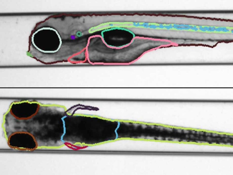
Decorative Image for HI Collaboration Identifying structural features of zebrafishes, using Semantic Segmentation
 

Identifying structural features of zebrafishes, using Semantic Segmentation

Zebrafish have a certain genetic similarity to humans and vertebrates. Particularly, the use of embryos is attractive due to the small scale screening capacity. Furthermore, similar to genuine cellular in vitro approaches, zebrafish embryos are considered as alternatives to animal testing. Therefore, they play a fundamental role in the detection of environmental and human health […]
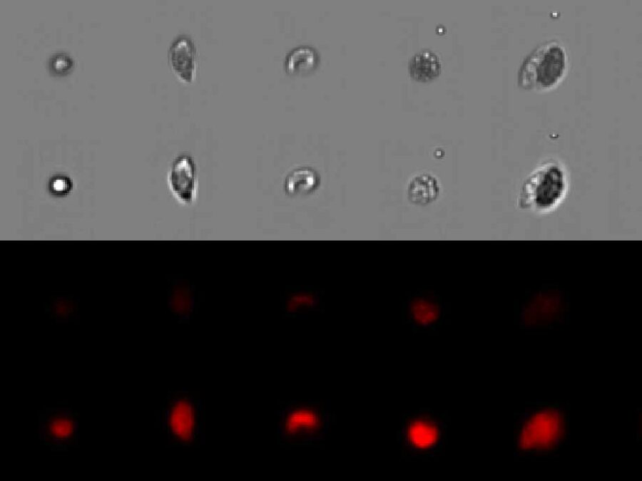
Decorative Image for HI Collaboration Automated Analysis of Evolutionary Experiments of Phytoplankton
 

Automated Analysis of Evolutionary Experiments of Phytoplankton

There is a strong interest in understanding community assembly and dynamics. Experimental approaches using phytoplankton have proven to be extremely insightful to unravel underlying biological processes. Imaging flow cytometry is an emerging method becoming more and more popular in different fields. It allows us to capture changes within a community or population in a more […]
 

Understanding lung diseases and optimizing their treatment

Understanding the distribution of nanoparticles after inhalation can lead to the discovery of novel, more effective drug delivering techniques. To this end we develop AI-based methods for semantic segmentation of the airways in non-dissected whole murine lungs, imaged with light sheet fluorescence microscopy. The analysis of airway properties in diseased lungs will furthermore shed light […]