FOMIA
A Foundation Model for Microscopy Image Analysis
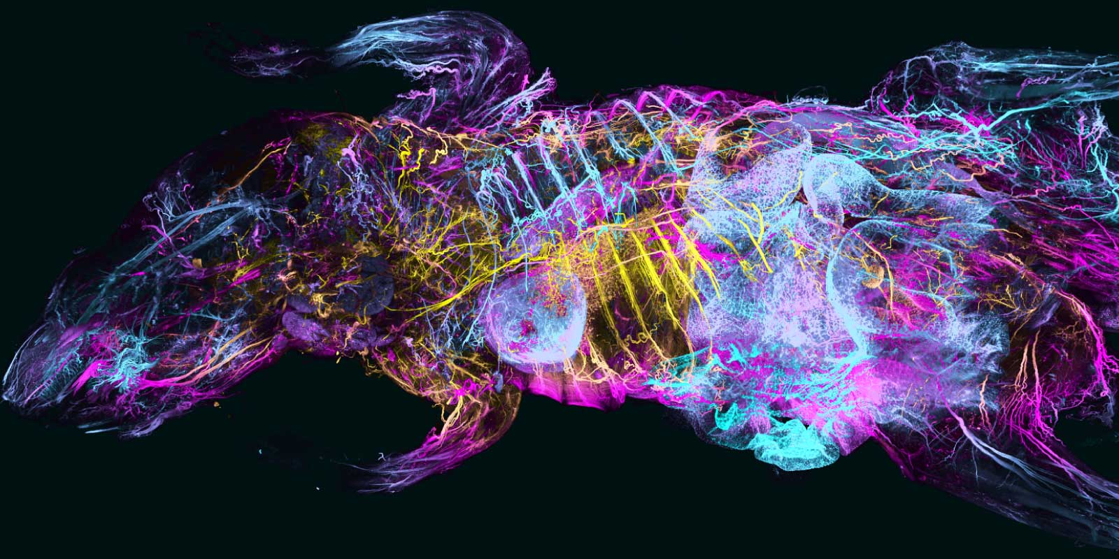
FOMIA aims to develop a powerful foundation model for the analysis of 2D and 3D fluorescence microscopy images.
We will harness a unique library of 3D light sheet microscopy images of whole mice and human organs, as well as a wide variety of 2D and 3D images from different sample types, organisms, microscopy modalities, and staining methods. Using this diverse data, we will employ self-supervised learning to train a robust foundation model capable of recognizing biological structures across scales.
The model will be fine-tuned for specific segmentation and classification tasks using minimal annotated data, significantly reducing the need for manual labeling. Its performance will be evaluated using public benchmark datasets, established methods, and newly generated data from our institutes.
To ensure practical applicability, the project includes a Co-Design collaboration with Deep Piction GmbH in Munich. All trained models will be made publicly available.
Other projects
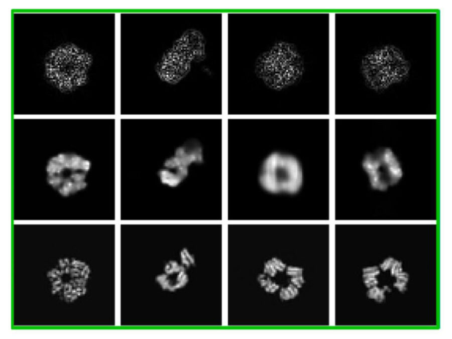
cryoFocal
3D Reconstruction from defocused cryo-EM images
This project explores how defocused images recorded with an electron microscope can be used to reconstruct the 3D structure of molecules inside cells. This method aims to enable faster and more cost-effective structural analysis of molecules to accelerate understanding of their functions and to design drugs against them.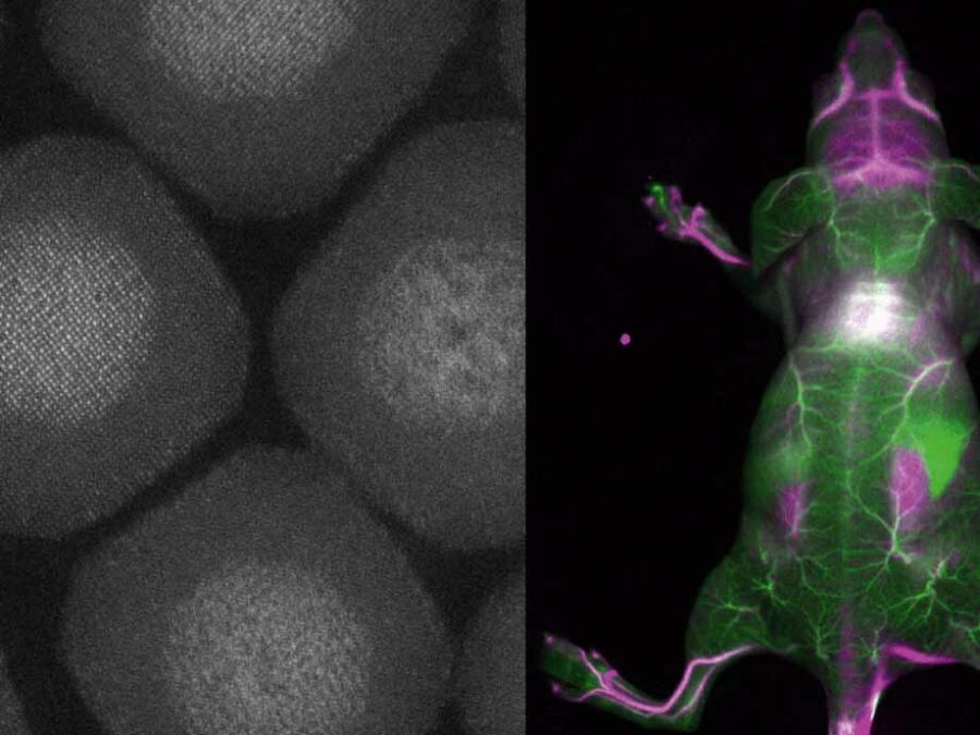
BENIGN
Biocompatible and Efficient Nanocrystals for Shortwave Infrared Imaging
The BENIGN project aims to enable non-invasive molecular imaging with cellular resolution in vivo at depths of several millimeters. This will be achieved using light from the shortwave infrared (SWIR) range (1000-2000 nm), which has less scattering and autofluorescence compared to the visible and near-infrared spectral range. Bright and targeted imaging agents are needed to fully exploit this range. The project will develop a new approach using lanthanide-based core-shell structures that emit light in the 1500-2000 nm range.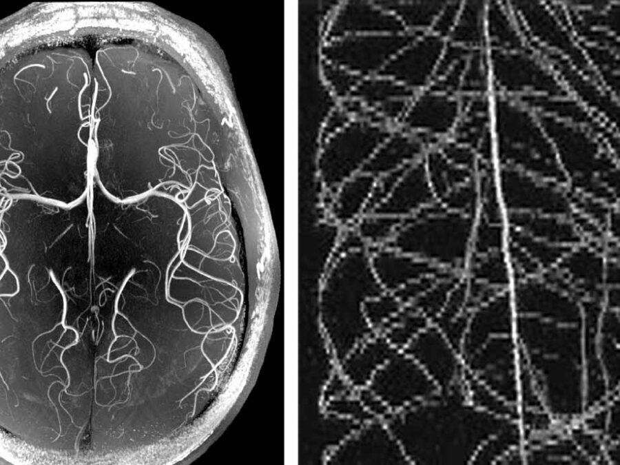
HighLine
High Image Quality for Lines in MRI: From Roots to Angiograms
MR images of roots and vessels are very similar: both display thin, line-like objects. The aim of the project is to increase image quality of both kind of MR data by exploiting their similarity. HighLine aims at obtaining high quality images in reduced scan time to lower patient burden and increase patient and plant throughput by adapting state-of-the-art 3D image enhancement methods, and developing new deep-learning based methods.