CLARITY
CineMR-guided ML-driven Breathing Models for Adaptive Radiotherapy
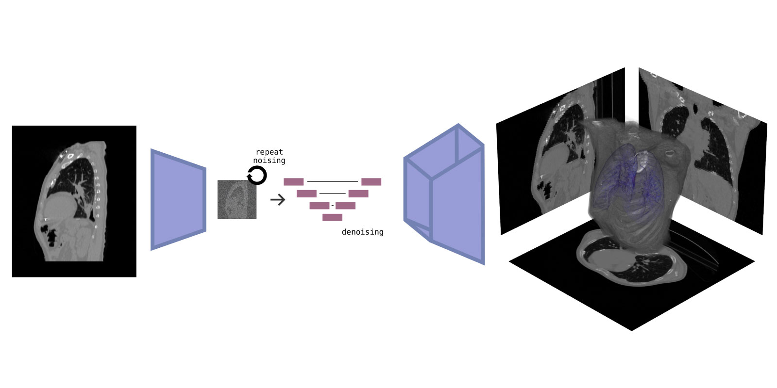
The integration of 2D cine-MR imaging in lung cancer radiation therapy has significantly improved treatment of moving targets, allowing for reduced safety margins and dose escalation. This approach has led to better overall survival rates for lung cancer patients. However, available tracking methods do not account for the full range of anatomical trajectories during breathing, missing out-of-plane motions of critical structures. This limitation makes current gating techniques insufficient for tumors located near the hilar or mediastinal regions, where dose escalation would result in irradiation of adjacent moving aortico-pulmonary structures, causing unacceptable toxicity.
To safely escalate doses in ultra-central lung tumors, fast image vision and generative extrapolation beyond integrated MR imaging is essential.
Detecting of out-of-plane moving organs at risk in the high-dose area would allow triggering of an irradiation interlock in the clinical gating scenario. However, this detection has to be robust, artifact-resistant, and fast. Within this project, we aim to develop novel algorithms to detect subtle movements in 2D cine scans. This will help recognize out-of-frame organ movements, improving the accuracy of radiation doses to cancerous tissue while sparing critical organs.
Long-term, improving 2D temporal anomaly detection is insufficient. Advances in MR-guided particle therapy, where the interplay effect between motion and beam scanning causes more severe dosimetric consequences, require full 4D motion information. Dose computation and re-optimization need full beam path anatomy estimates. As real-time 4D imaging is not feasible, we will advance existing generative AI algorithms to three dimensions, allowing volumetric anatomy extrapolation and continuous tumor motion tracking.
Other projects
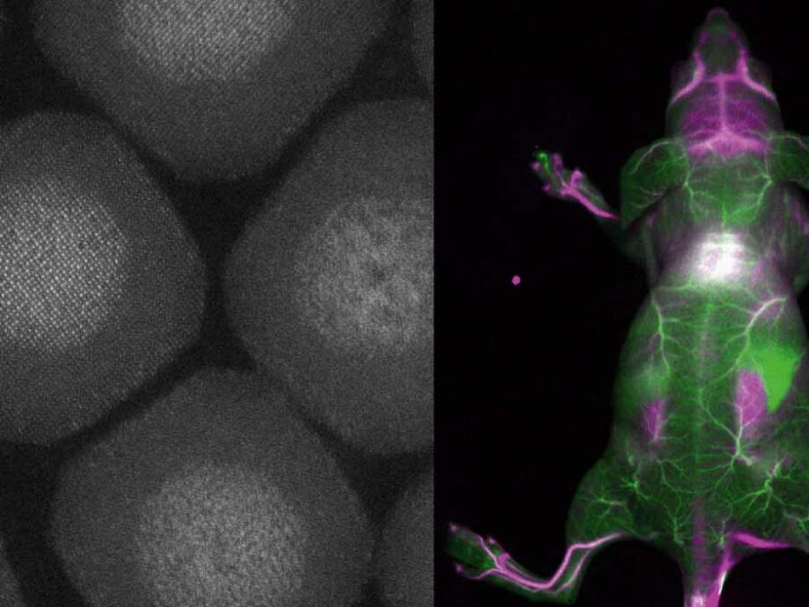
BENIGN
Biocompatible and Efficient Nanocrystals for Shortwave Infrared Imaging
The BENIGN project aims to enable non-invasive molecular imaging with cellular resolution in vivo at depths of several millimeters. This will be achieved using light from the shortwave infrared (SWIR) range (1000-2000 nm), which has less scattering and autofluorescence compared to the visible and near-infrared spectral range. Bright and targeted imaging agents are needed to fully exploit this range. The project will develop a new approach using lanthanide-based core-shell structures that emit light in the 1500-2000 nm range.
TerraByte-DNN2Sim
On the trail of the mystery of the laws of calving
Researchers still face a mystery when it comes to the laws by which glaciers calve. This project aims to use satellite imagery, artificial intelligence, mathematical optimisation and a new data processing pipeline to track the movements of glacier fronts in Antarctica to get closer to solving the mystery.
Hyper 3D-AI
Artificial Intelligence for 3D multimodal point cloud classification
The aim is to develop an artificial intelligence that can achieve the fusion of two-dimensional data with three-dimensional information. Based on this, the software would simultaneously be able to recognise image characteristics as well as the spatial relationships between different objects.