UCS
Ultra Content Screening for Clinical Diagnostics and Deep Phenotyping

When asked to describe their research, Dr. Oliver Bannach and Dr. Thomas Rockel like to show a picture: it portrays London’s famous Tower Bridge with its two iconic towers, but the left half of the picture is rendered as an outline of the bridge, reduced to three colours, while the right half of the picture is a full colour photo in sharp detail. “You can imagine the difference between previous imaging methods and our new technology to be like this,” the two scientists from Forschungszentrum Jülich explain. “We want to increase the informative value of images by a long way.”
The project aims to develop technology that can automate the screening of single cells from the human body. Based on the existing method called ultra content screening technology (UCS), a new system will now be developed that can fully automate the examination of a blood sample. This involves staining individual components of the cells with fluorescent antibodies and photographing them under a special microscope.

This is then repeated for another component of the cell, and then another – gradually creating a detailed view inside the cell, layer by layer. They will first be doing this for 30 selected biomarkers in tumour cells and bone marrow cells from breast cancer and leukaemia patients. After that, the cells of those patients will be examined for any additional striking commonalities.
Pipetting robots and other high-end tools will perform the analyses automatically. The imaging and data analysis software required for interpreting the resulting data will also have to be developed in the project.
Other projects
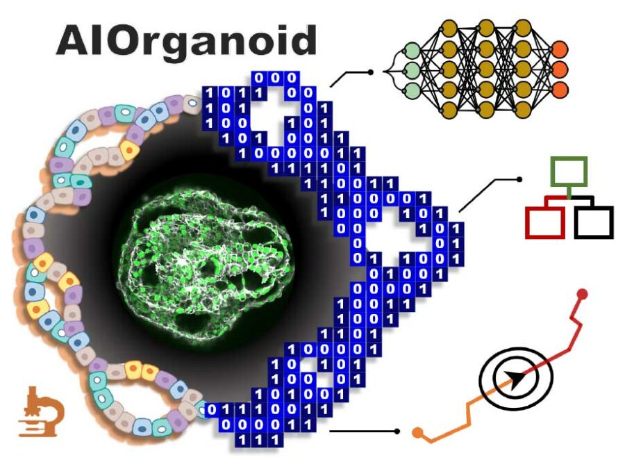
AIOrganoid
Artificial Intelligence Assisted-Imaging for Creating High-yield, High-fidelity Human Lung Organoid
AIOrganoid will apply cutting-edge imaging techniques and develop novel AI-based solutions to facilitate human lung organoid formation with high yield and fidelity, bridging the gap between cell biology and computational imaging.CLARITY
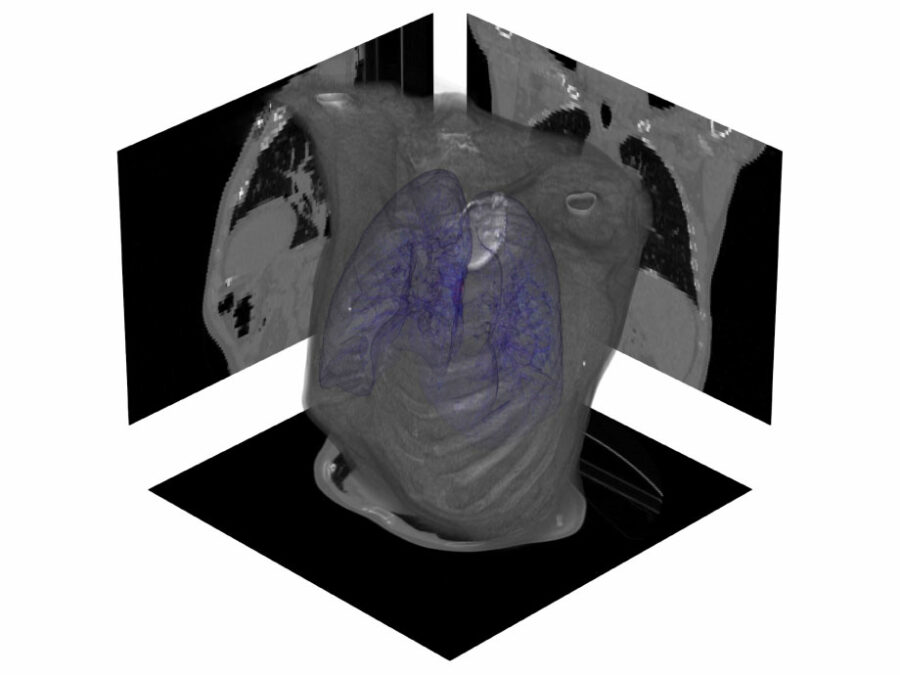
CineMR-guided ML-driven Breathing Models for Adaptive Radiotherapy
Dose-escalated radiotherapy of lung cancers requires precise monitoring of lesions and nearby organs at risk. Current methods are able to track ultra-central lesions but neglect their deforming vicinity, risking unacceptable toxicity to aortico-pulmonary structures. AI-based anomaly detection and generative AI models can address both requirements in real-time.HighLine
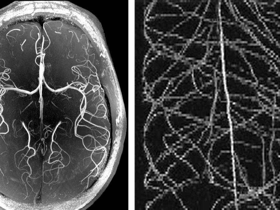
High Image Quality for Lines in MRI: From Roots to Angiograms
MR images of roots and vessels are very similar: both display thin, line-like objects. The aim of the project is to increase image quality of both kind of MR data by exploiting their similarity. HighLine aims at obtaining high quality images in reduced scan time to lower patient burden and increase patient and plant throughput by adapting state-of-the-art 3D image enhancement methods, and developing new deep-learning based methods.

