
Best Scientific Image Contest 2025
In its fifth edition, the Best Scientific Image Contest once again brought together the Helmholtz community’s outstanding imaging talents. 104 fascinating images were submitted – explore the top 21 in our virtual gallery!
Gallery
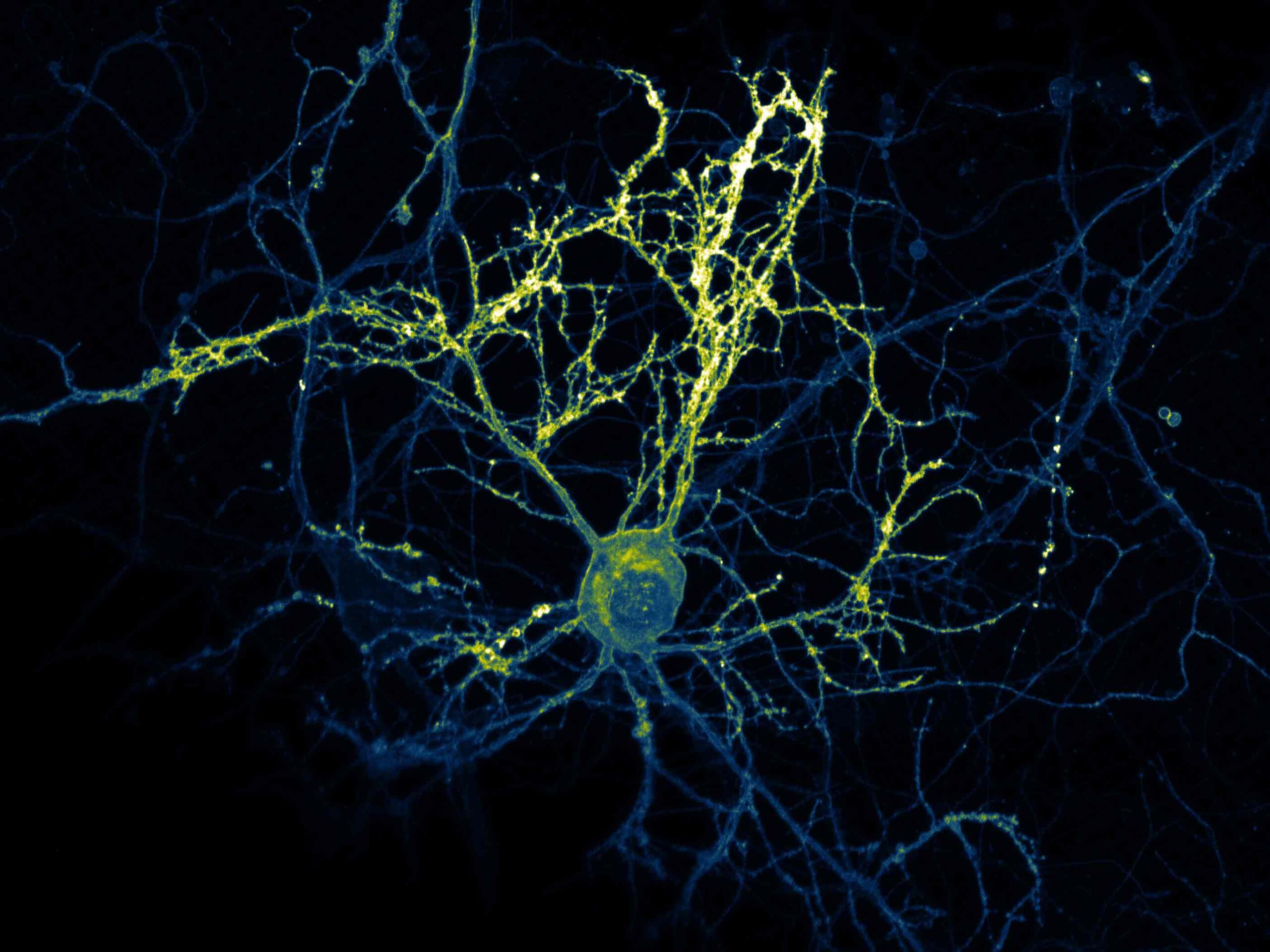
Excitation (1st place Jury Award)
Jana Kroll, MDC
The image shows neurons labeled with a sensor for glutamate release. Variations in fluorescence intensity correspond to signal strength, providing insights into neuronal activity and connectivity. The neurons were optogenetically stimulated and immediately shock-frozen by cryofixation at the moment of neurotransmitter release. The cryo-confocal image not only confirms successful stimulation but also serves as a guide for cryo-electron tomography by correlating fluorescence and electron microscopy data. This enables the activity of individual synapses to be linked to neuronal morphology, offering a detailed understanding of synaptic signaling within a single neuron. Captured as a z-stack using a Leica SP8 cryo-confocal microscope operated at cryogenic temperature (below −170 °C).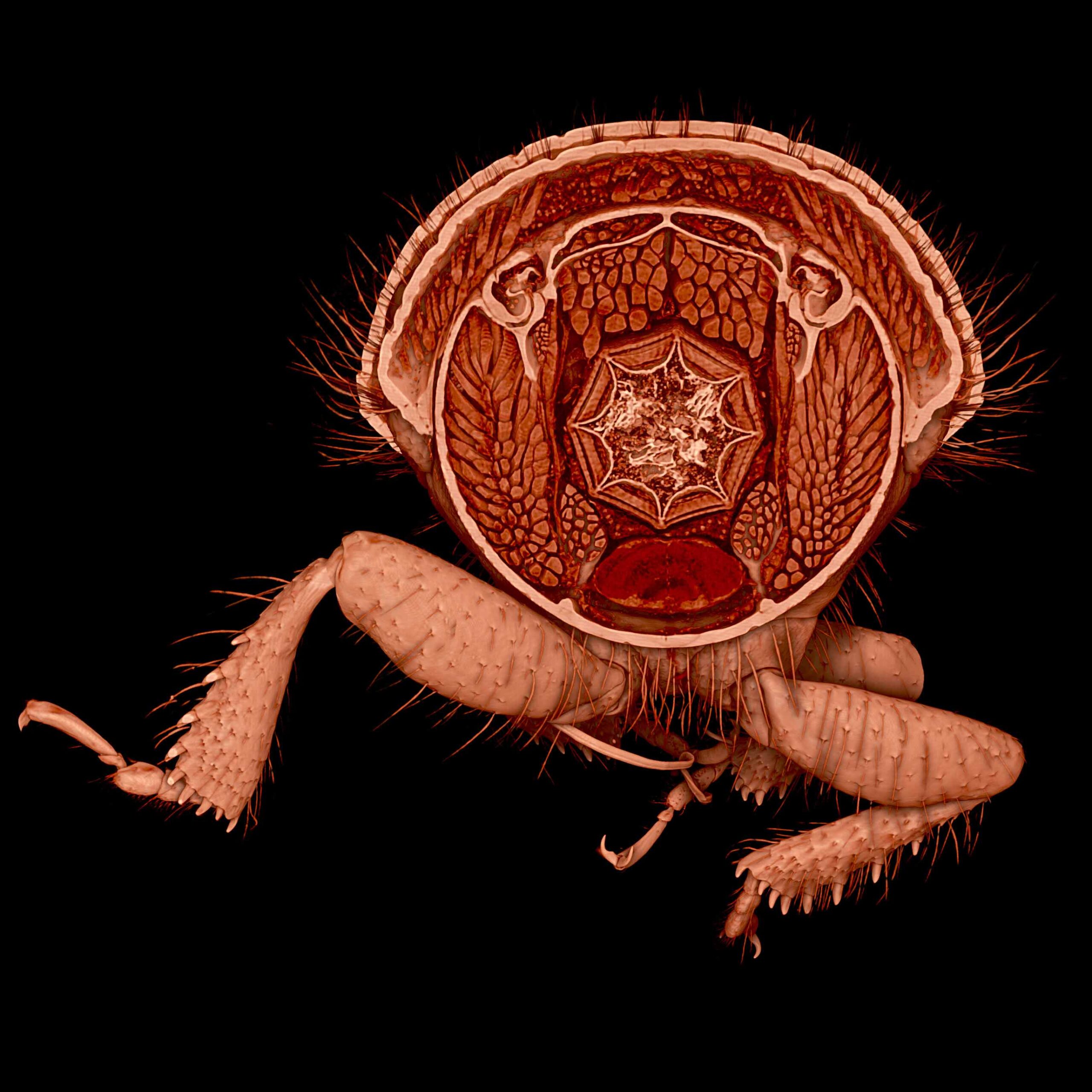
A Bark Beetle’s Stellar Gut (2nd place Jury Award, 3rd place Public Choice Award, 1st Place Participants' Choice Award (shared))
Jenny Hein, Thomas van de Kamp, KIT
A virtual cut through a high-resolution 3D scan of a bark beetle reveals its intricate internal structures, including a striking star-shaped foregut, highlighting the unseen marvels of the microcosmos. Insects, Earth’s most diverse group of animals, play essential roles in ecosystems, agriculture, and biomimetics. Understanding their full morphology in 3D is vital for exploring key adaptations and biodiversity. Large-scale digitization of specimens, like the one shown here, enables correlations between internal anatomy, molecular data, and environmental context, deepening biological insight. Captured using a filtered polychromatic beam at the high-throughput microtomography station of the IMAGE beamline at KIT Light Source. The software Drishti was used to create a volume rendering, digitally cut to highlight internal features and refined using Adobe Photoshop.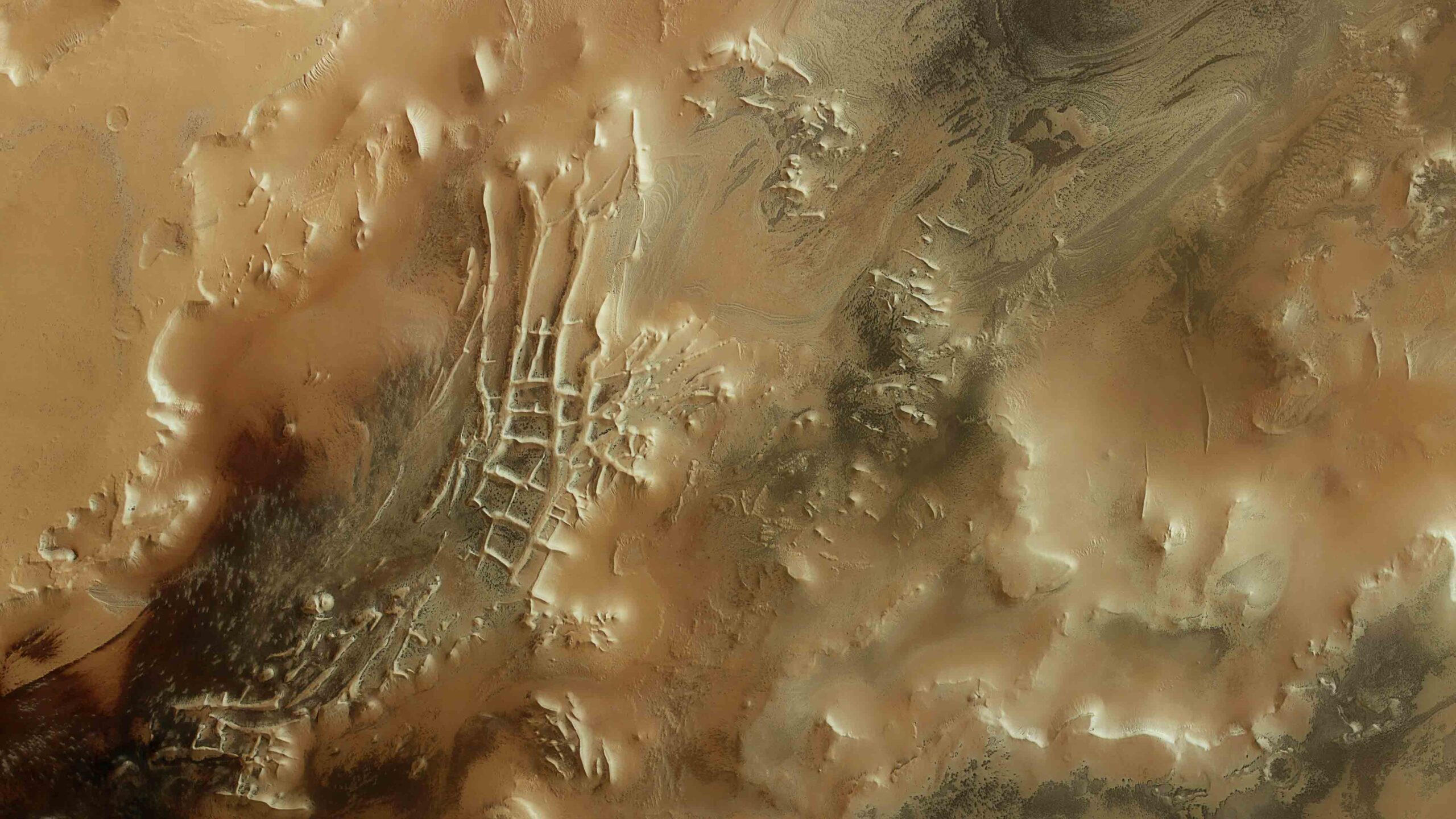
"Inca City" on Mars (3rd place Jury Award)
Daniela Tirsch, DLR, Björn Schreiner, FU Berlin
A stunning view of Angustus Labyrinthus, a maze-like ridge system nicknamed “Inca City” near Mars’ south pole, this image offers crucial insights into the planet’s polar geology, revealing processes that have shaped its climate and surface over time. It displays intricate, intersecting ridges surrounded by polar layered deposits. Scientists are investigating whether these formations resulted from tectonic faulting, glacial flow, or sublimation of subsurface ice, each scenario providing clues about Mars’ dynamic history. Captured using orbital imaging from about 350 km above the surface by the High Resolution Stereo Camera (HRSC) onboard ESA’s Mars Express.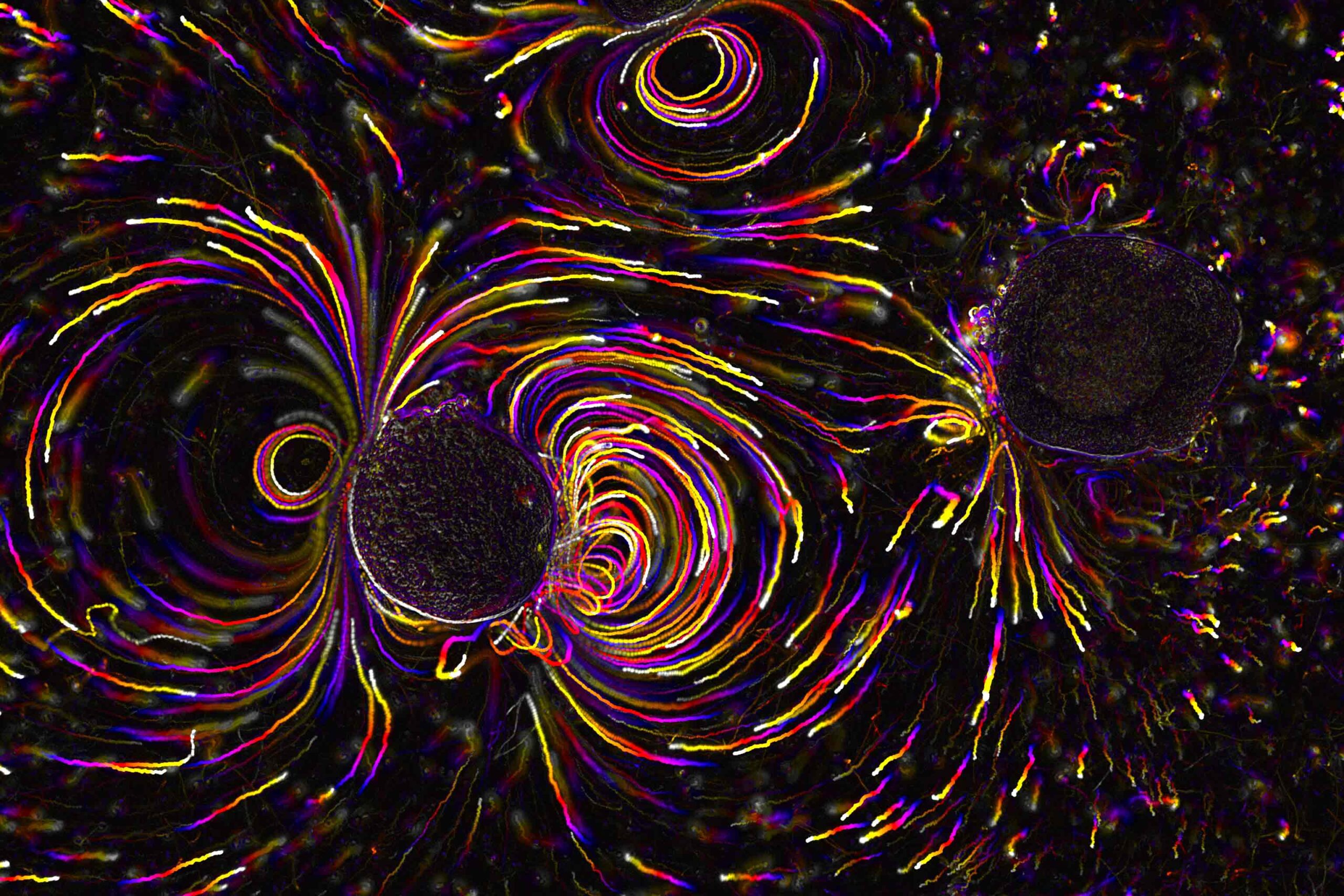
Starfish Puts Motion Back in the Ocean (1st place Public Choice Award)
Rafael Deliz-Aguirre, MDC
This image shows starfish embryos generating tiny currents that push them together to form crystal-like arrangements. The living embryo on the left actively sculpts its fluid environment, in contrast to the motionless, dead control on the right. Their microscopic tentacles create currents that mirror Earth’s magnetic shield visually and mathematically, and led to equations that might also apply to black hole plasmas and herds. These structured flows offer insights into active matter physics and chiral materials. Starfish embryos provide a cost-effective model to study self-organizing systems and develop mathematical models of Stokes flow, where viscosity dominates. Vortex-like patterns in these flows resemble planetary magnetic fields, revealing deep connections between microscopic and celestial physics. Captured using brightfield microscopy with fluorescent beads, then temporally color-coded to visualize dynamic flow patterns.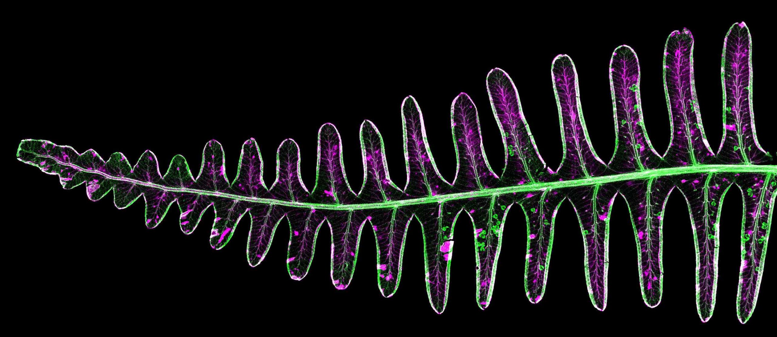
Rare Earth Element Fern (2nd place Public Choice Award)
Kathryn Spiers, Dennis Brückner, DESY, Antony van der Ent, Wageningen University, Léo Goudard, Université de Lorraine, Germinal Rouhan, Muséum national d’Histoire naturelle, Paris
This image shows where the rare earth element yttrium is concentrated in a frond of the fern Dicranopteris linearis. Different colours represent the spatial distribution of elements, with bromine standing out in the spherical sporangia and yttrium forming striking patterns across the leaf tissue. During the analysis of the historical Paris Herbarium collection, this fern was identified as having the highest concentration of rare earth elements (REEs) ever found in a plant. Certain ferns possess the unusual ability to hyperaccumulate REEs, making them promising candidates for phytomining, an emerging technology that uses plants to extract critical minerals from low-grade sources and industrial waste. Understanding the molecular mechanisms behind hyperaccumulation could help develop sustainable alternatives to traditional mining. Captured at beamline P06 of PETRA III, DESY, using X-ray fluorescence microscopy.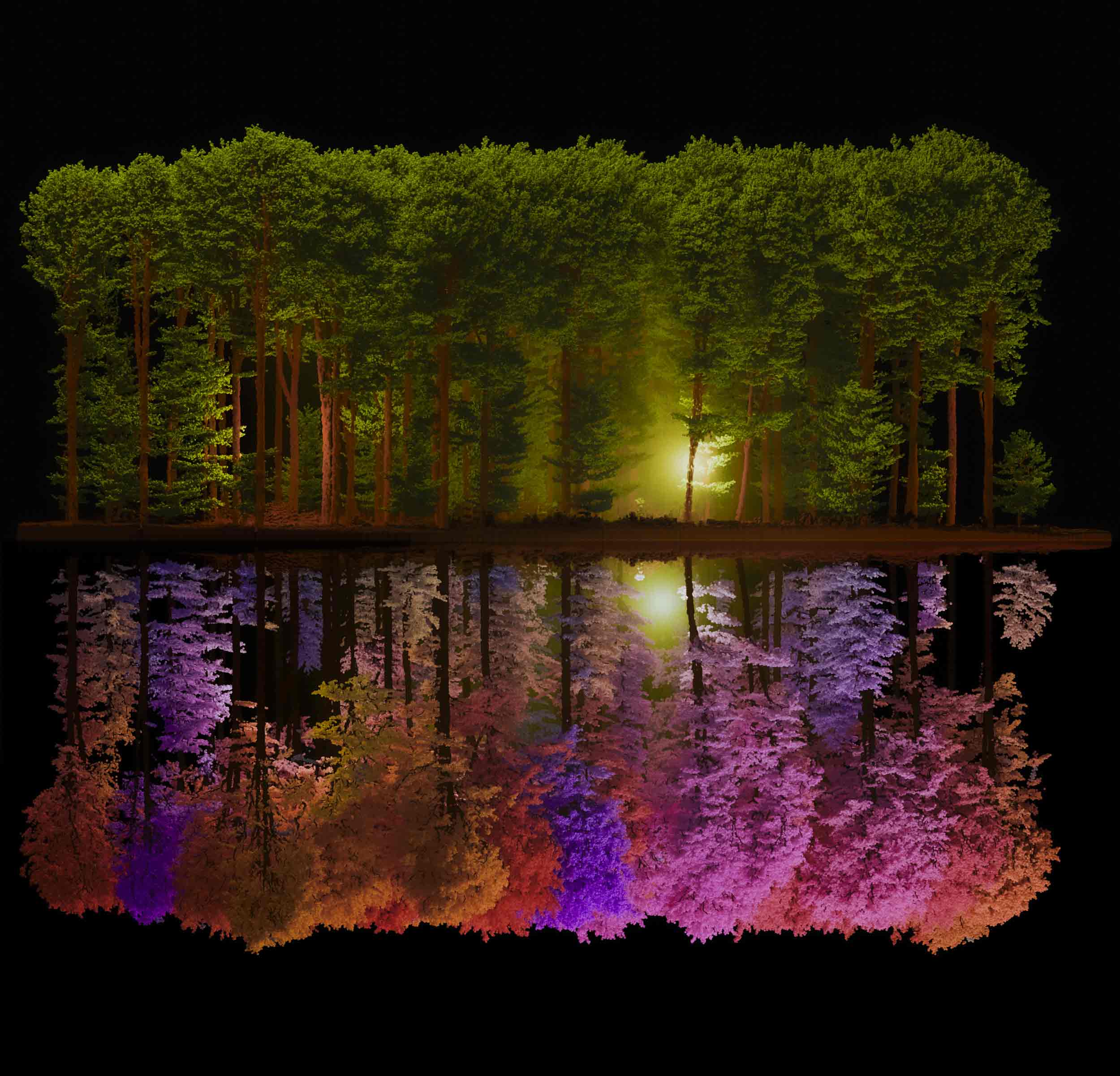
Digital Forest / Digital Twin (1st Place Participants' Choice Award (shared))
Aldino Rizaldy, Sam Thiele, Sharad Kumar Gupta, HZDR
Forests are key in the climate and biodiversity crises. This light detecting and ranging point cloud uses machine learning and graph theory to aid monitoring, conservation, and better management. The data shown here forms a digital twin of a real forest in Hohes Holz (Saxony-Anhalt), an accurate, three-dimensional representation that enables detailed analysis of forest structure and health. Advanced algorithms identify individual trees and their components, supporting applications such as biomass estimation, carbon stock assessment, and species mapping. Captured using a Riegl VUX-120 airborne laser scanner mounted on a Velos V3 POR drone and rendered using Blender software to create a detailed visualization.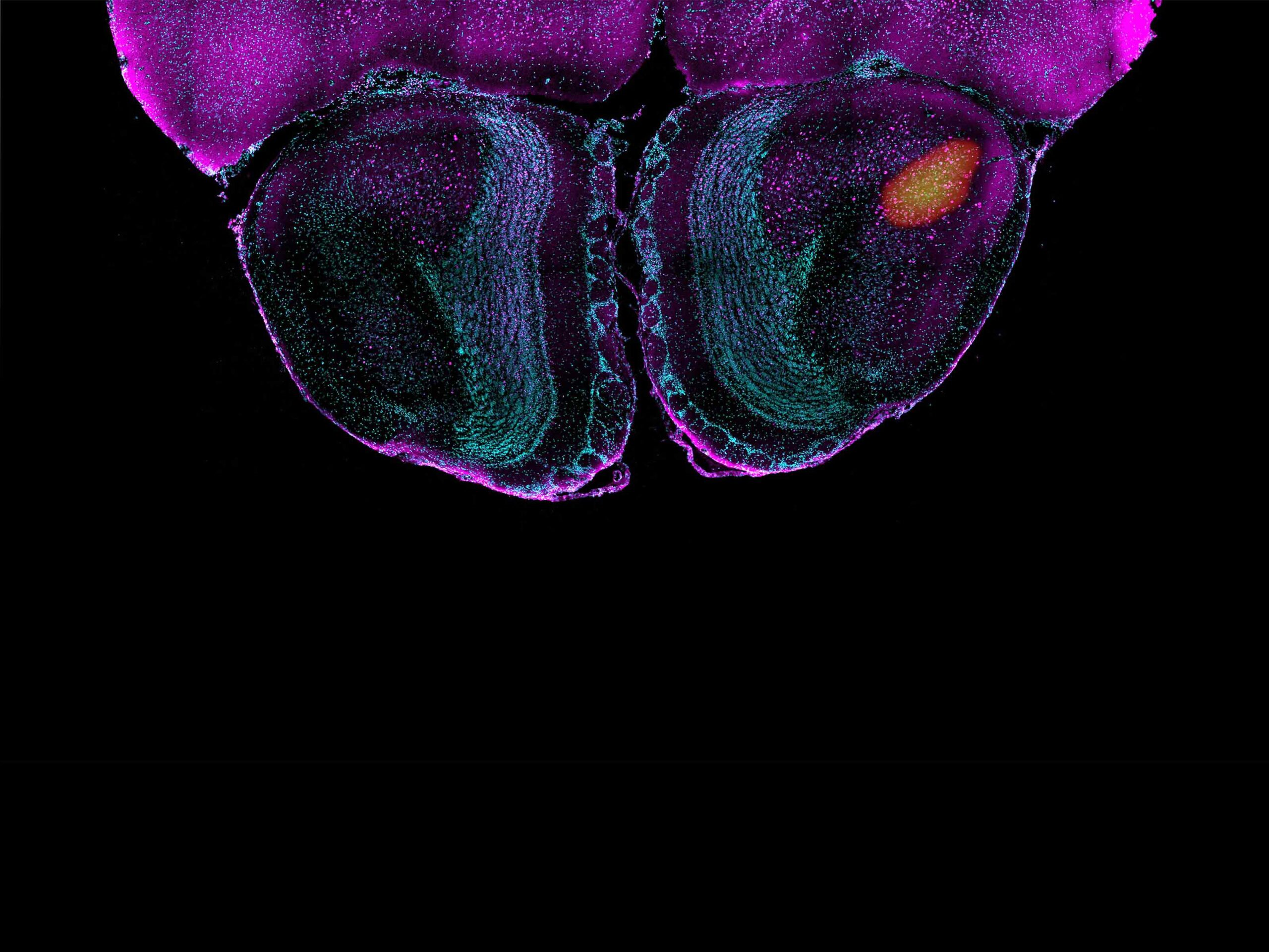
Filming the Smelling Brain (3rd Place Participants' Choice Award)
Athanasios Balomenos, Wenhan Luo, Gary R. Lewin, MDC
This image combines functional ultrasound imaging and high-resolution confocal microscopy to illustrate how specific stimuli activate the smell center in anesthetized mice. The highlighted brain region is the anterior olfactory nucleus (AON), a key part of the olfactory cortex involved in processing smells. Despite its prominent position in the olfactory pathway, the AON remains poorly understood. By imaging its activation in real time, researchers are beginning to uncover previously unknown functions of this region. The results suggest that the AON responds selectively to certain stimuli, offering fresh insights into its role in olfactory perception. Captured using the novel functional ultrasound imaging (fUSI) and a spinning disk confocal microscope.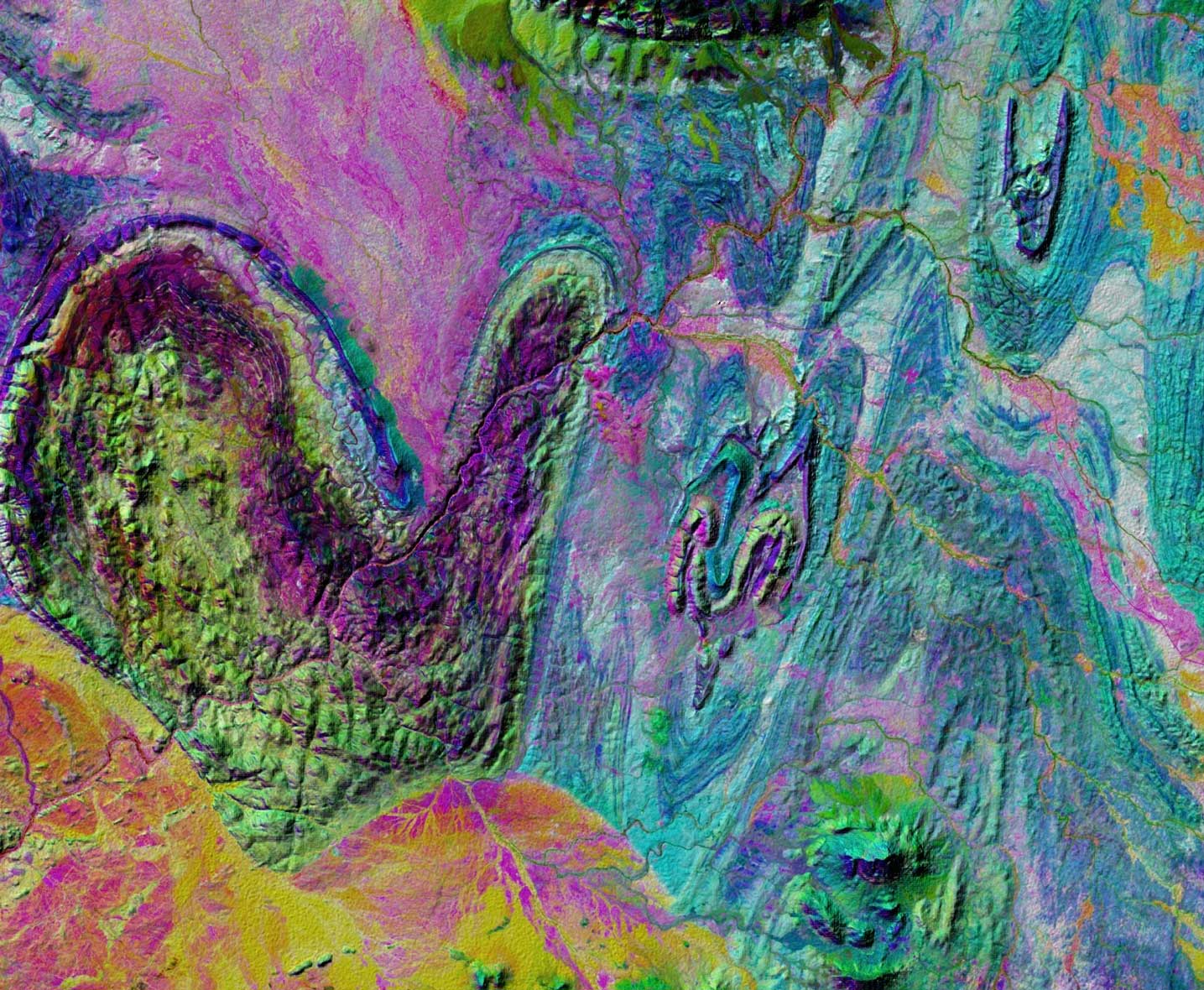
Thumbs-Up from Earth
René Booysen, Richard Gloaguen, HZDR
This image shows how satellite data support sustainable mineral exploration for green technology. It merges spectral and topographic data to reveal the geological complexity of a 1200 km² area in northern Namibia. The vibrant colors result from a Principal Component Analysis (PCA) applied to Sentinel-2 multispectral data, enhancing subtle spectral differences between rock types, while a PALSAR-derived elevation model shows terrain features such as folds and faults. Visible are a mitten-shaped structure and a circular magmatic intrusion now mined for raw materials. By detecting infrared light beyond human vision, scientists can identify rock types and mineral-rich zones with minimal environmental impact. Captured using Sentinel-2 multispectral satellite data (10 m resolution) and a Digital Elevation Model from PALSAR Synthetic Aperture Radar.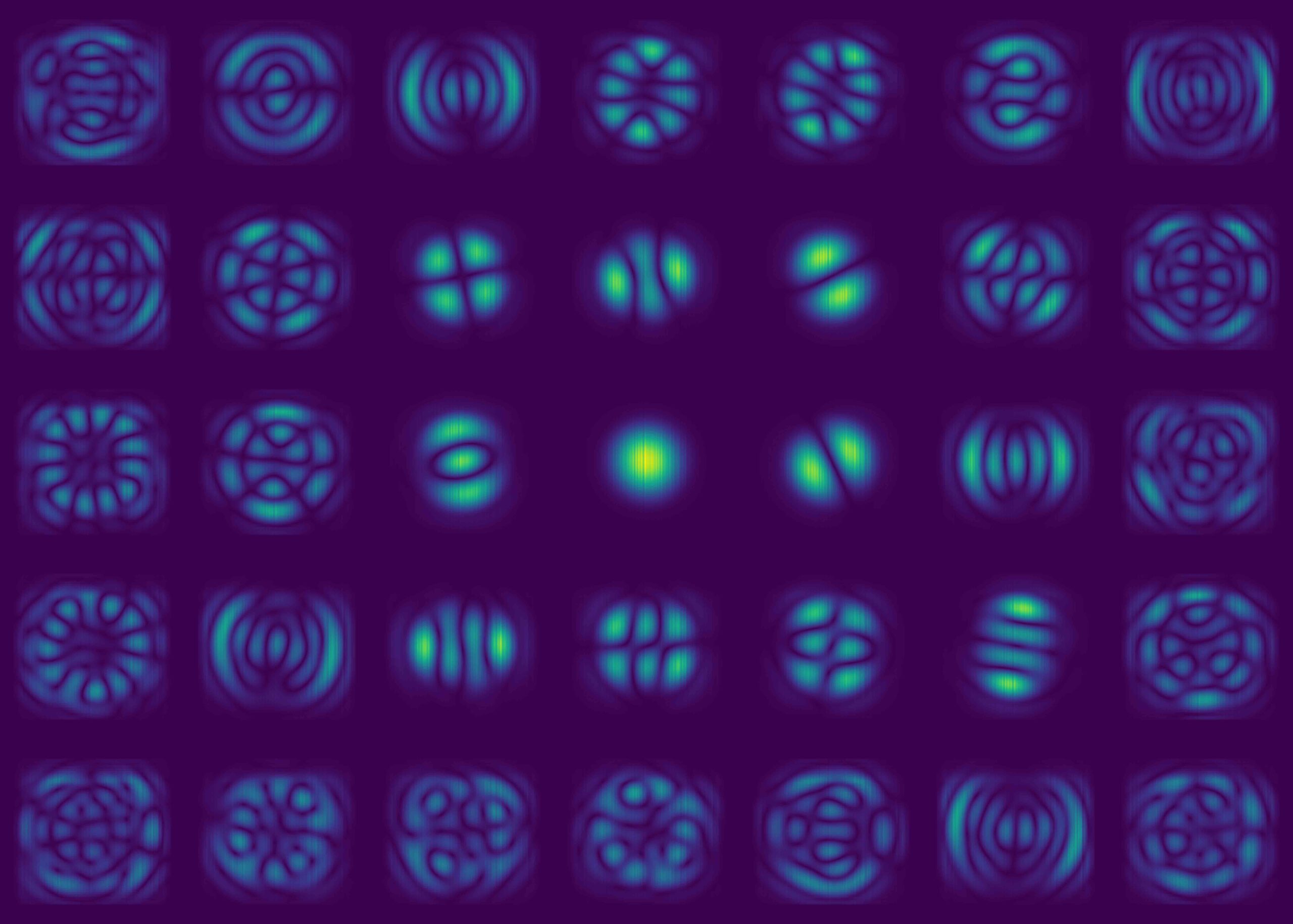
The Perfect Round Beam
Martin Seyrich, DESY
The image shows simulated contributions to the beam in an X-ray microscope. The round shape and high intensity of these contributions predict world-leading performance for the future Ultimate X-ray Microscope PETRA IV at DESY in Hamburg. The simulation currently serves to guide and validate the optical design of the beamline and define key parameters, such as the required quality of mirror surfaces. It also supports the development of X-ray optics and novel coherent imaging methods. Looking ahead, the goal is to transform the simulation into a digital twin of the beamline that enables scientists and users to plan and optimize experiments, maximizing scientific output from the limited and valuable beam time. Captured from a multi-electron wave-propagation simulation of a PETRA IV X-ray Nanoprobe beamline, using adapted elements of the SRW and XRT simulation codes.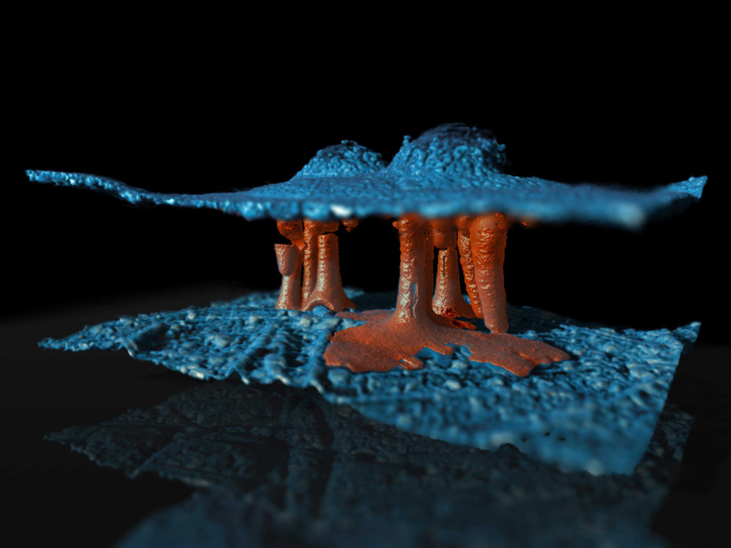
Tiny Threads, Big Impact
Christine Heume, FZJ
This 3D image shows a novel degradation mechanism inside a membrane electrode assembly: filaments (red) connect anode and cathode (blue), causing an electrical short circuit. Such images offer valuable insights into failure processes in PEM water electrolyzers. The three-dimensional view reveals how degradation evolves over time. By tracking filament growth, researchers can identify weak points and improve system design and durability. These insights are essential for boosting the performance and reliability of PEMWE systems, which are key to scaling green hydrogen production. Captured using X-ray computed tomography with the ZEISS Xradia 620 Versa Micro-CT system, the image was acquired at a voxel size of 534 nm. Coloring and visualization were performed using Dragonfly Pro.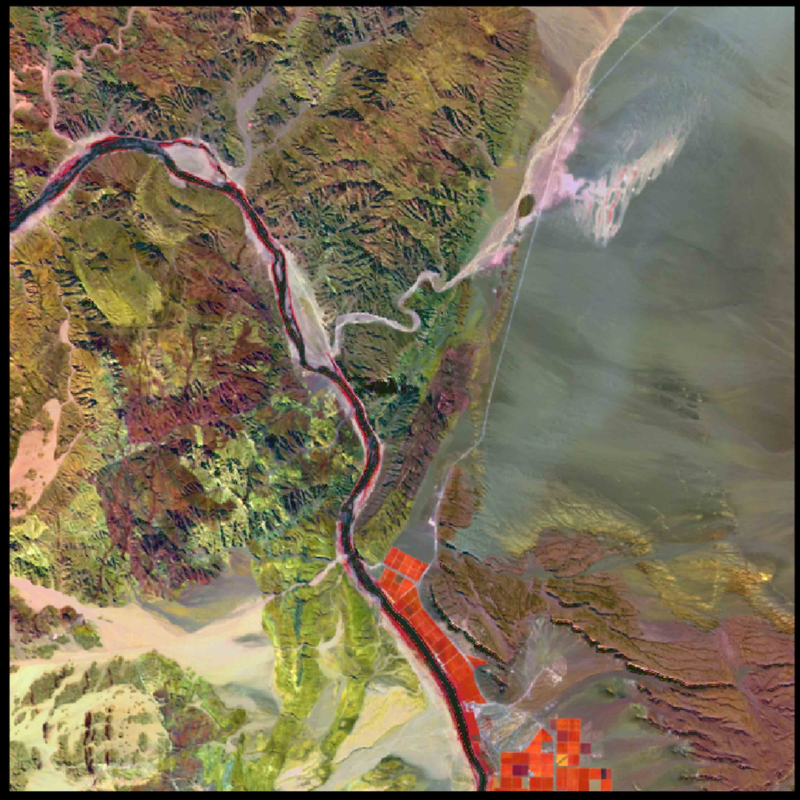
Hyperspectral Data Uncovering the Unseen
Rupsa Chakraborty, HZDR
A bird’s-eye view of a rocky region in Marinkas Quellen, Namibia, rich in critical raw materials, is enhanced with hyperspectral data, revealing subtle geological variations and offering valuable insights for mineral exploration. The raw image was captured by the EnMAP sensor, developed by DLR, and overlaid with results from a data decomposition analysis to visualize spectral variability. This region holds resources vital for technologies ranging from renewable energy to electronics. Spaceborne hyperspectral data, such as from EnMAP, allows high-resolution mineral mapping over large, remote areas without disturbing the environment. Combined with field validation and machine learning, it enhances the precision of resource assessments. Captured using a spaceborne hyperspectral imaging sensor.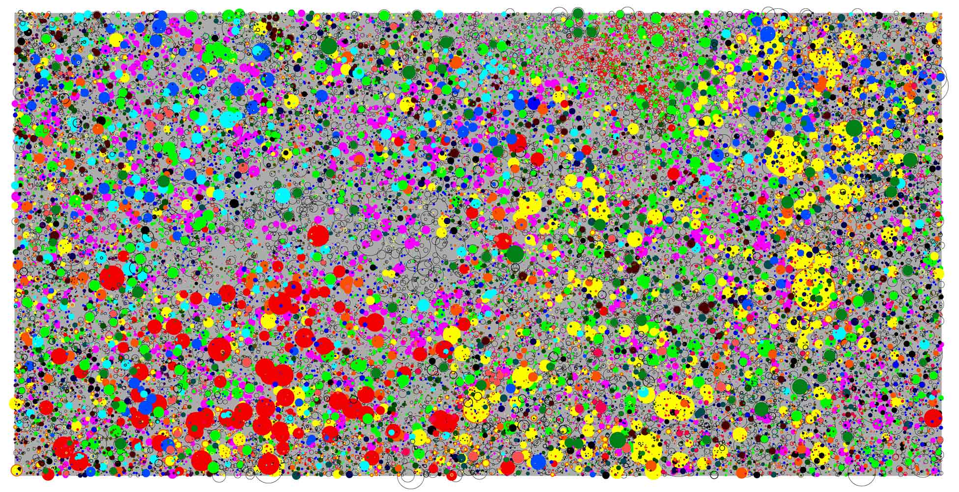
Tropical Forest on Barro Colorado Island
Thorsten Wiegand, UFZ
This image shows the spatial position, species (color), and size of 87,570 trees in a 1000 m × 500 m section of tropical forest on Barro Colorado Island in the Panama Canal. Each tree with a diameter at breast height >2.5 cm is shown as a disk scaled proportionally to its stem diameter. The 28 most abundant species are highlighted in color, layered from most to least abundant. This visual representation raises a central question in ecology: how can so many tree species coexist in such small areas? The spatial arrangement reflects ecological processes emerging from interactions between individual trees and their environment. To explore this, mathematical and individual-based simulation models are combined with high-quality spatial forest inventory data. Comparing observed and simulated patterns helps identify mechanisms of coexistence and test ecological theories. Captured using SigmaPlot.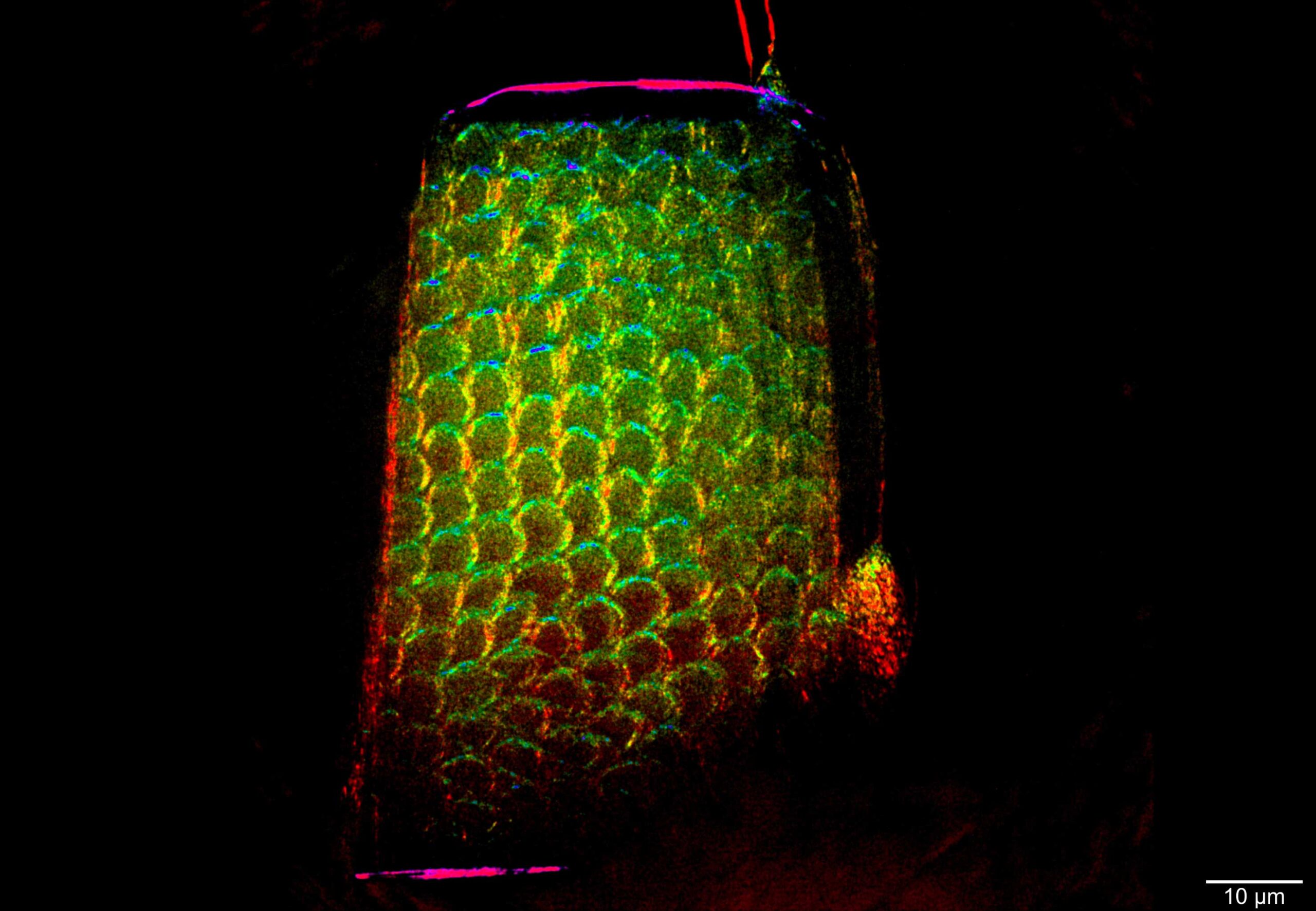
A Tooth Fairy Secret
Sami Wirtensohn, HEREON
The first application of directional nano dark-field imaging shows the enamel of a human tooth. The color encodes the crystalline orientation, derived from how X-rays scatter in different directions. This imaging technique visualizes internal structures based solely on scattered intensity, producing a unique type of contrast. Since scatterers can lie below the system’s spatial resolution, dark-field imaging can uncover features that remain invisible to traditional transmission or phase-contrast techniques. Crucially, this method reveals both the strength and orientation of scatterers within each pixel, enabling the detection of aligned nanostructures, such as fibers or crystals, beyond the optical limit. This deepens our understanding of material properties and could enable future full-field nano-tensor tomography. Captured using dark-field transmission X-ray microscopy to visualize SAXS intensity in a full-field image.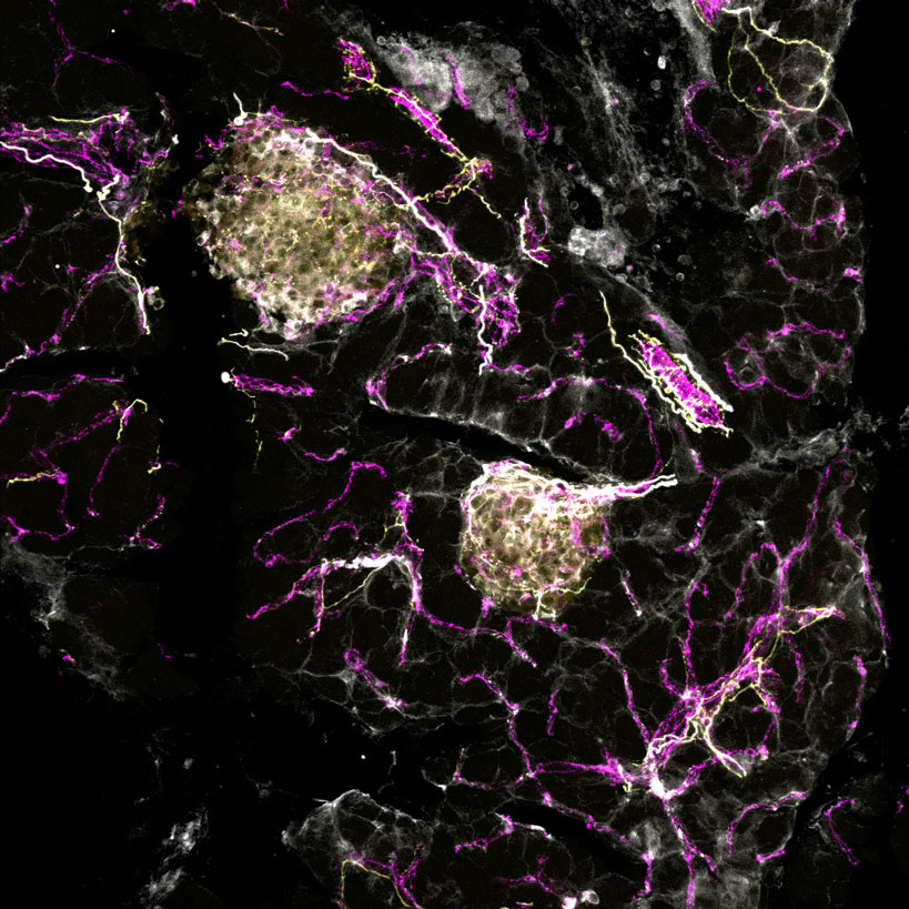
Well-connected
Andreas Müller, HM
Pancreatic islets, blood vessels, and the nervous system must work closely together to regulate our blood glucose levels. This fluorescence image shows how nerve cells (yellow) and blood vessels (magenta) interact with the islets of Langerhans (white) to ensure proper insulin secretion. By visualizing these structures, researchers aim to gain a more holistic view of insulin regulation by simultaneously examining the different cell types interacting in the pancreas. This integrated approach also helps improve our understanding of diabetes. Captured using confocal microscopy.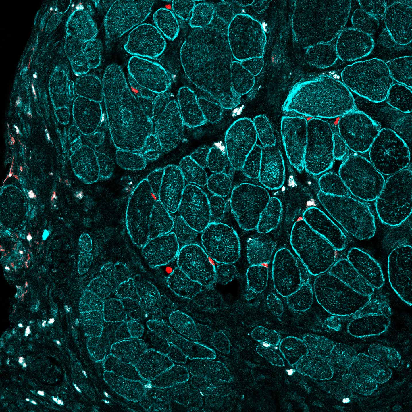
Healthy Muscle After Gene Editing
Andreas Marg, MDC
This image shows regenerated muscle tissue in humanized mutant mice following the transplantation of gene-corrected muscle stem cells. The newly formed fibers express dysferlin (cyan), while Pax7-positive satellite cells (red) mark the restored regenerative capacity. These findings demonstrate that gene-edited cells can significantly improve muscle integrity in diseased tissue. The repair was achieved using CRISPR/Cas9 gene editing tools and offers promising prospects for treating dysferlin-deficiency, one of several currently untreatable muscular dystrophies. The image was taken three weeks after transplantation from a 6 µm thick section of the tibialis anterior muscle, stained for dysferlin and Pax7. Captured with a Zeiss LSM 900 and a 40x/0.95 objective (image size: 355 µm × 355 µm).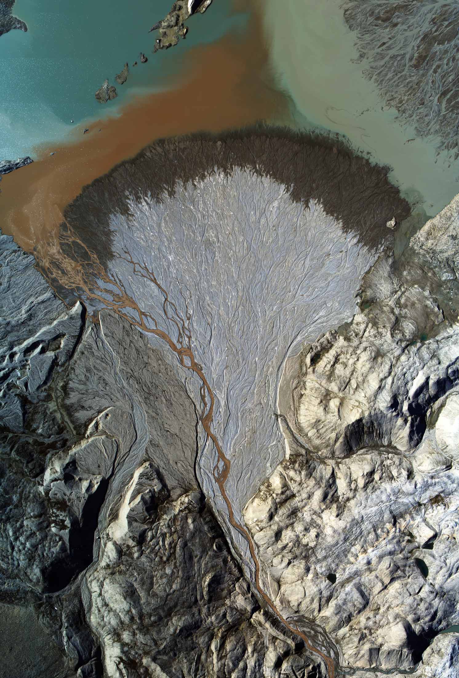
Beauty of Arctic Glacial Decline
Tilman Bucher, Ernst Hauber, DLR
This aerial image shows a delta in a proglacial lake fed by meltwater from the rapidly retreating Uvêrsbreen glacier on Svalbard. The vivid landscape illustrates the beauty and fragility of the Arctic environment in the face of climate change. As glaciers recede and permafrost degrades, landforms evolve at unprecedented rates. Researchers aim to identify and quantitatively monitor the rates of these geomorphological processes in a rapidly changing glacial and permafrost environment and link them to global climate change. Captured using the DLR MACS modular aerial camera system onboard AWI's Polar-6 aircraft from a flight altitude of 800 meters.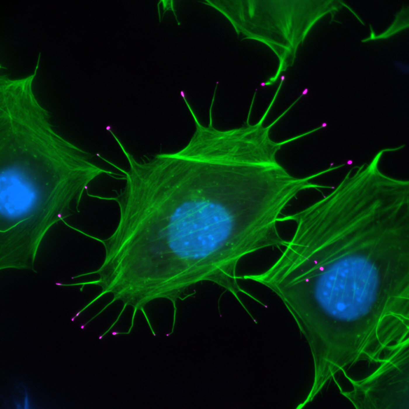
Myosin X at the Tips of Filopodia
Anika Steffen, HZI
This fluorescence image shows spreading mouse cells with finger-like protrusions called filopodia, which contain actin filaments (green); DAPI staining visualizes cell nuclei (blue). The cell in the middle overexpresses Myosin X (magenta). These cells were genetically modified to delete the small GTPase Rac1, a protein well-known to drive actin-regulated plasma membrane protrusions called lamellipodia. Without Rac1, the cells rely entirely on filopodia to explore their environment. By visualizing the precise localization of Myosin X during these dynamic processes, researchers can better understand how motor proteins and actin filaments cooperate to drive membrane protrusion and cell movement. This image highlights the importance of individual molecular players in shaping cellular behavior. Captured using epifluorescence microscopy.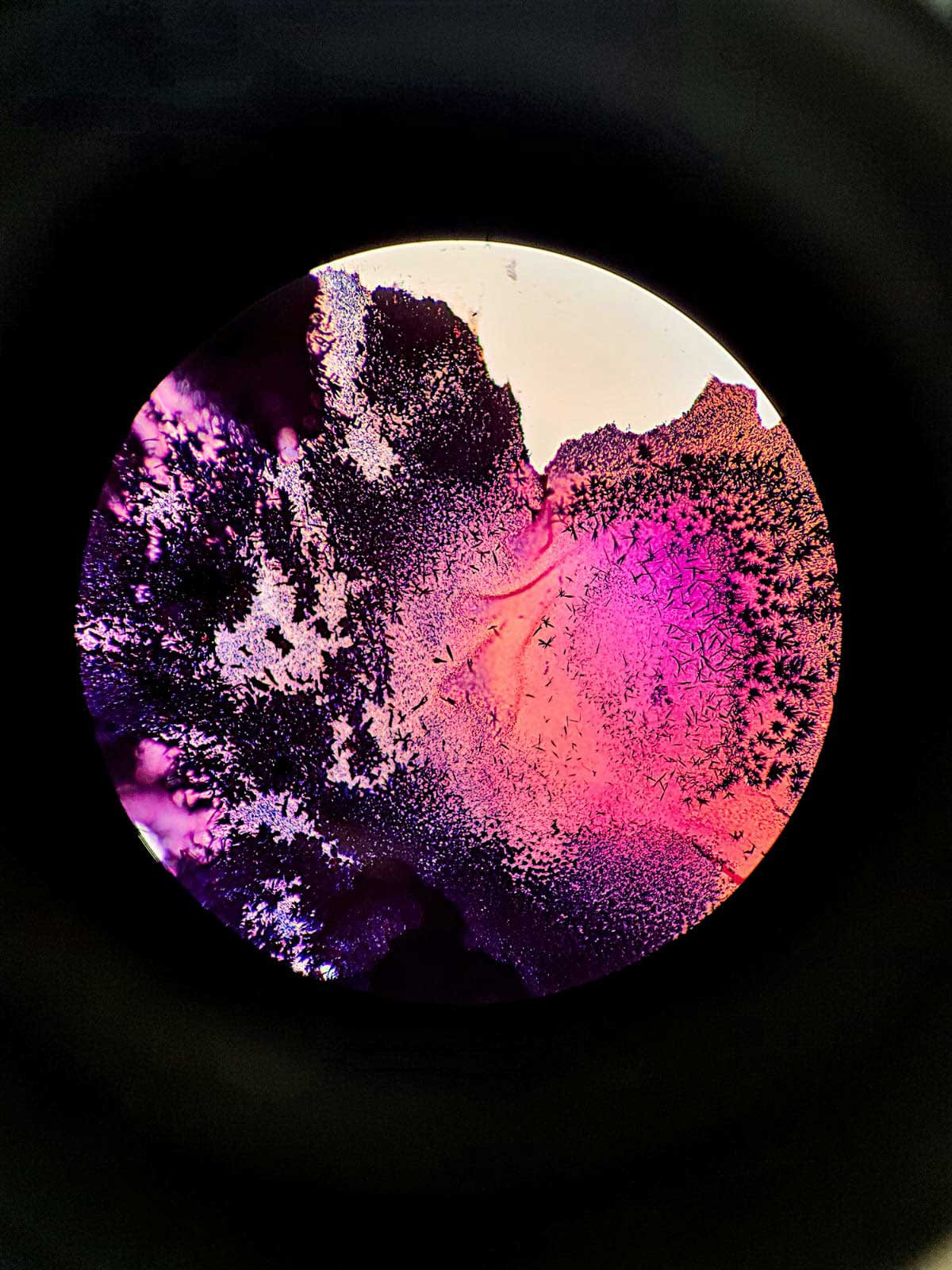
A Stunning Failure
Silas Beckmann, FZJ
A brief glimpse of failure, invisible to the naked eye, yet revealing its full splendor under the lens of a simple microscope. This image was taken by accident during a Gram staining procedure using crystal violet and safranin to assess bacterial behavior. Due to diffi culties with the Bunsen burner, the staining solution unexpectedly crystallized, producing intricate, colorful patterns. What was initially considered a failed experiment revealed an unexpected kind of beauty. Such moments remind us that even mistakes in the lab can off er new perspectives, spark curiosity, and uncover details that would otherwise remain unseen. Captured under a binocular using a common mobile phone.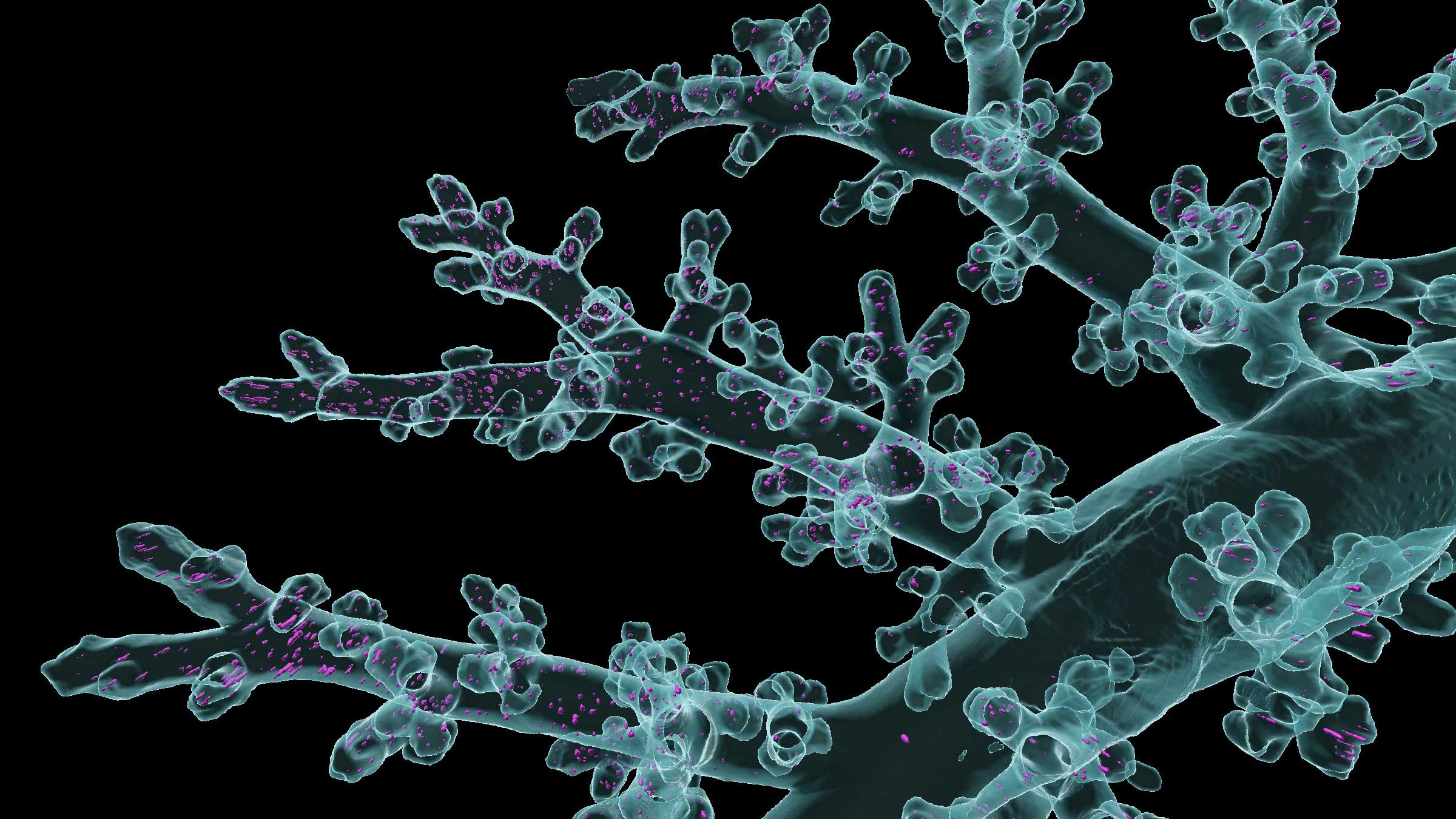
Aerosol Hotspots in Fine Airways
Lin Yang, HM
3D imaging and deep learning reveal how inhaled aerosols distribute within the complex structure of lung airways, showing preferential deposition in smaller branches and highlighting regional heterogeneity. The image offers a high-resolution view of large airways branching into distal regions, with visible hotspots of aerosol accumulation. These insights are critical for understanding how inhaled drugs reach different lung areas. Effective aerosol delivery is essential in respiratory therapy, where medications are administered via inhalers or nebulizers. Knowing how particles behave across airway generations supports the development of optimized drug formulations, improved inhaler designs, and targeted treatment strategies. Captured using light sheet fluorescence microscopy. Airway segmentation was performed using the LungVis 1.0 deep learning model, with aerosols and segmented airways rendered in magenta and cyan using Imaris.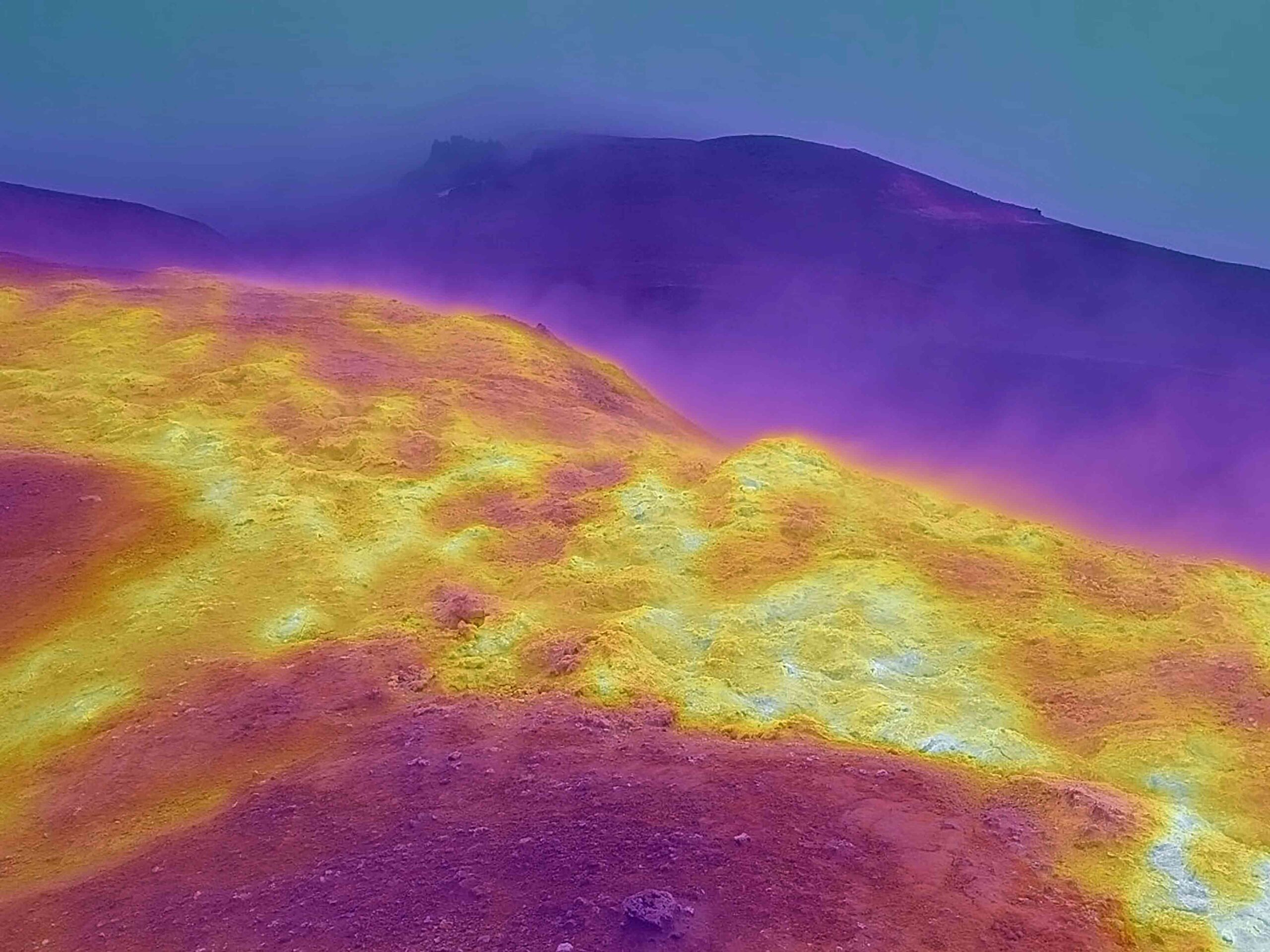
Geothermal Activity in Askja Caldera
Alina Shevchenko, GFZ
This infrared image of the geothermal field at the eastern wall of Askja caldera shows hotspot areas highlighted in brighter colors. Surface temperatures in this area reach up to 40 °C, while measurements at 20 cm depth indicate values as high as 70 °C. Infrared thermal imaging allows not only the measurement of surface temperatures but also the delineation of hotspot extent. These data can be compared with visible surface features such as sinkholes, cracks, and slumps to assess whether geothermal activity contributes to permafrost degradation and slope instability. Askja, one of Iceland’s largest calderas, is located in a geothermally active and permafrost-affected region, making it highly relevant for monitoring climate-related landscape changes. The image was captured in 2023 during fieldwork in Iceland using a FLIR radiometric thermal camera.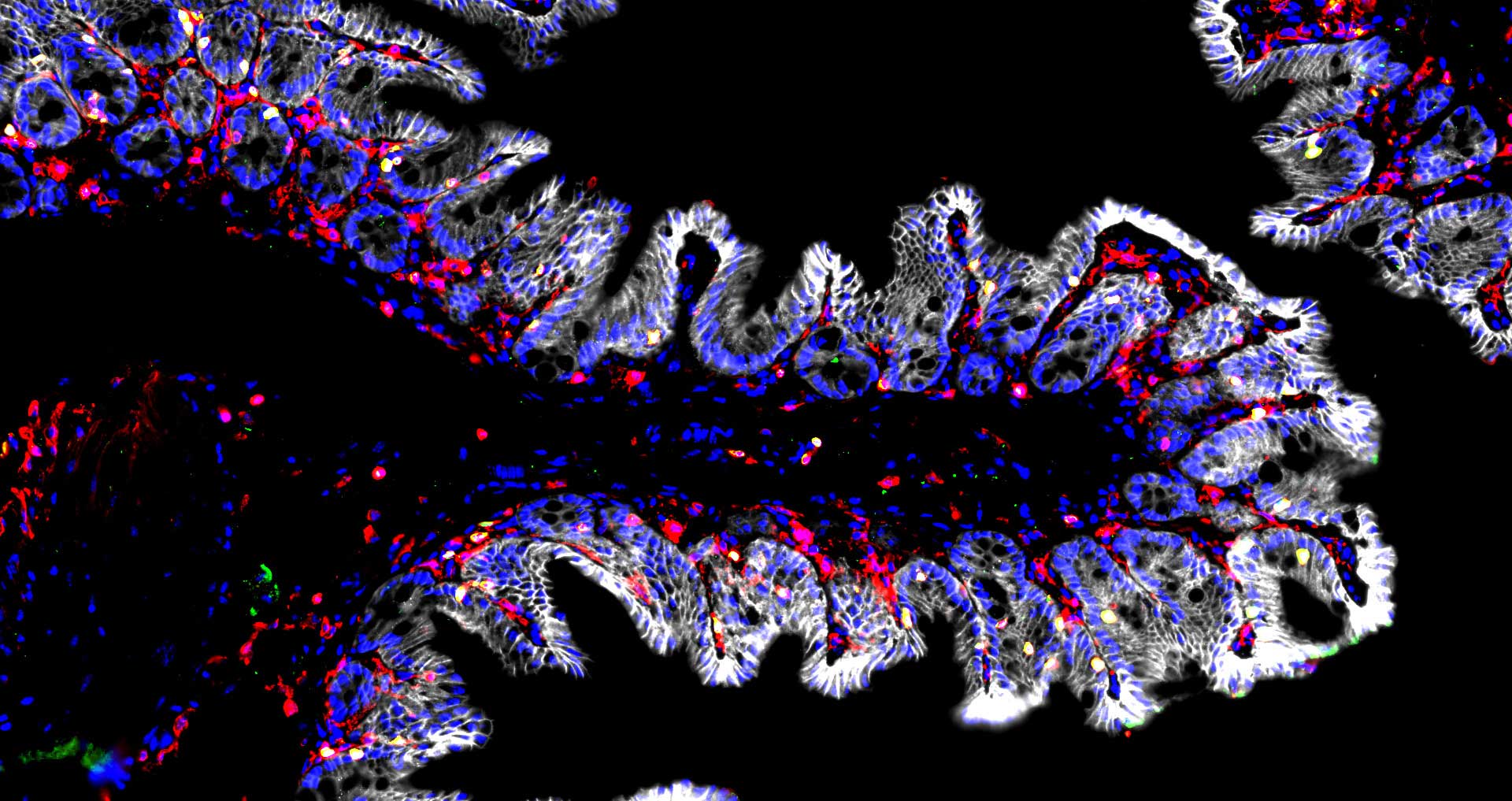
The Immune Landscape of the Gut
Ariana Rauch, MDC
This image shows the colon tissue in a mouse with a microbiome. Immune cells (red) and T-cells (yellow) are near or within the intestinal epithelium (grey), marking the border to the lumen and its microbes. This image investigates the location and distribution of immune cells and their dependence on the microbiome to identify immune subsets influenced by microbial presence. The findings aim to provide insights into alterations of immune cells in disease. Captured using fluorescence microscopy (3D Histech Slide Scanner) of antibody-stained PFA-fixed cryosections. Processing and coloring were performed in Fiji.