Best Scientific Image Contest 2023
In 2023 we called again for the Best Scientific Image. 162 high-quality, stunning images were submitted by the Helmholtz community. Discover the best 21 images in our virtual gallery!
Gallery

Lung health and immunity (1st Place, Jury Award)
Lin Yang, CPC/LHI, HMGU
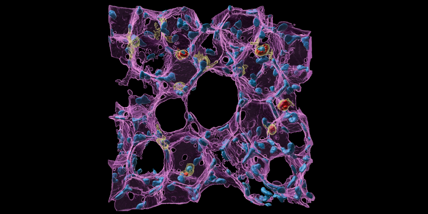
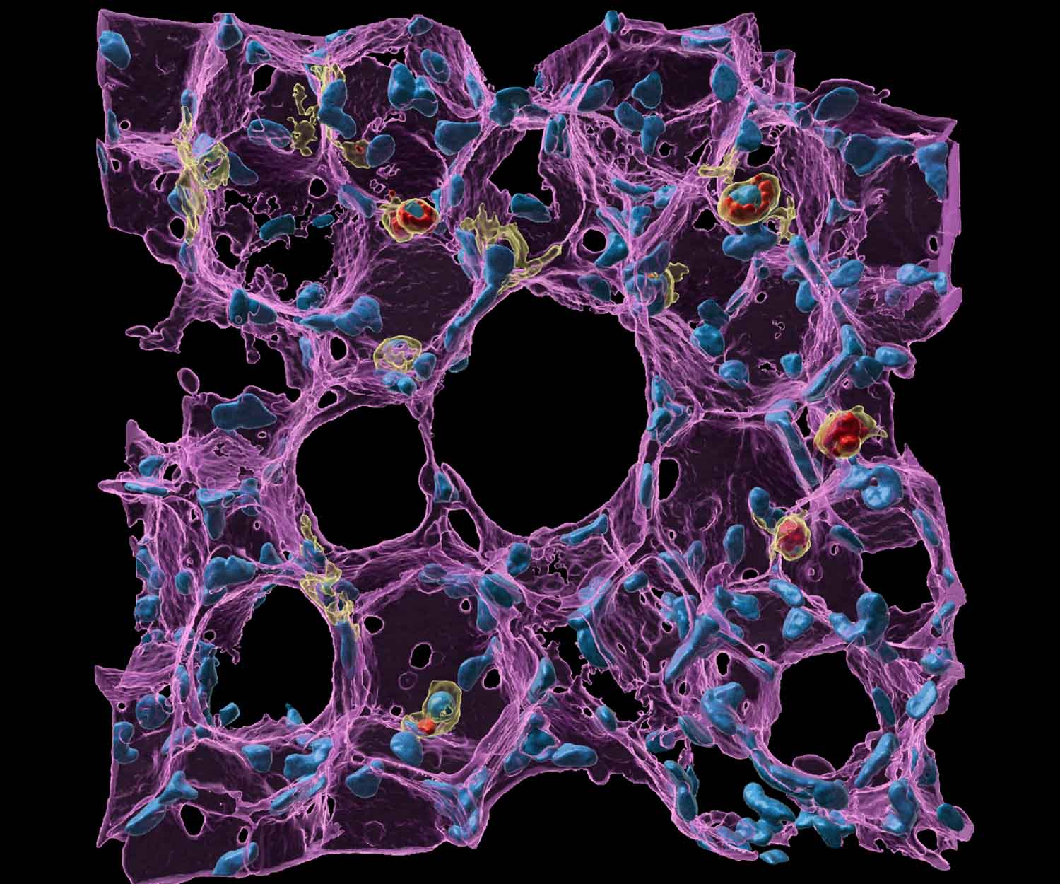
Lung macrophages remove inhaled toxins from lung alveoli, which is crucial for maintaining gas exchange and lung homeostasis. The image showcases a high-resolution, tiny but intricate lung alveolar architecture including interconnected lung alveoli, micro vessels, and capillaries (all in purple), as well as numerous locations of lung macrophages (yellow) engulfed with foreign pollutants (here fluorescent particles in red). The cell nuclei are in light blue. Lung macrophages actively patrol the alveoli, safeguarding against inhaled particles and maintaining homeostasis, while the visually striking pores of Kohn ensure air sac pressure balance and facilitate rapid immune response. This image, obtained with laser scanning confocal microscopy and created using Imaris, provides enriched information regarding the function of immune cells and magnificent lung alveolar geometry, inspiring us to prioritize care for lung health, due to direct environmental exposure.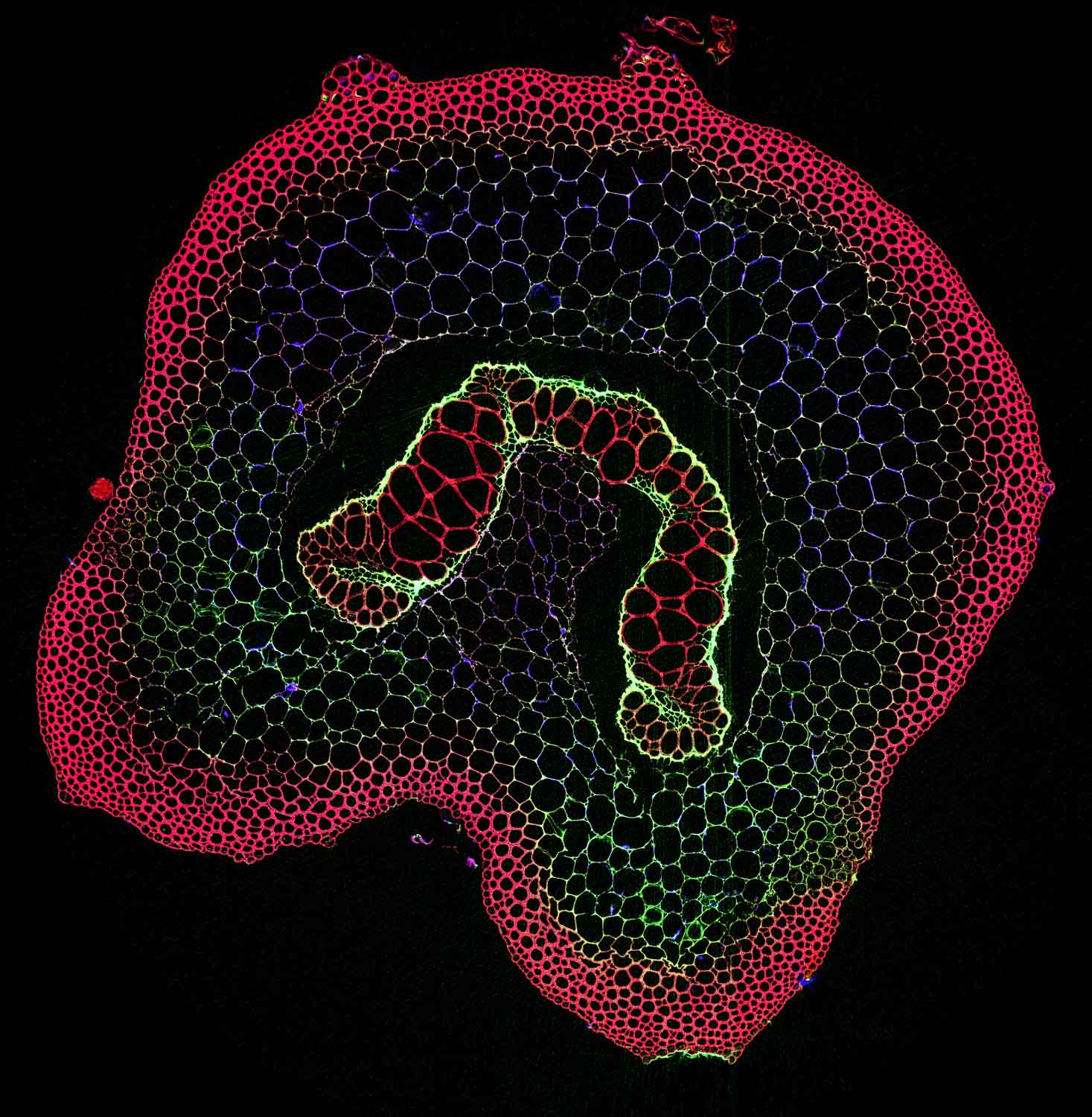

Arsenic fern (2nd Place, Jury Award)
Kathryn Spiers, Dennis Brückner, DESY & Antony van der Ent, Wageningen University & Research, The Netherlands
The tropical fern Pteris vittata has the exceptional ability to naturally accumulate arsenic into its living tissues. This image is a synchrotron micro-fluorescence computed tomographic reconstruction of a slice through the rachis (stem) of the fern. It was acquired at beamline P06 of DESY and shows arsenic in green, X-ray Compton in red and X-ray Absorption in blue color. It reveals that arsenic is localized in the endodermis and pericycle surrounding the vascular bundles. The elemental maps help to better understand the molecular and physiological mechanisms of arsenic tolerance in this fern. This information is used to study and improve its natural ability to accumulate arsenic for cleaning up contaminated soils.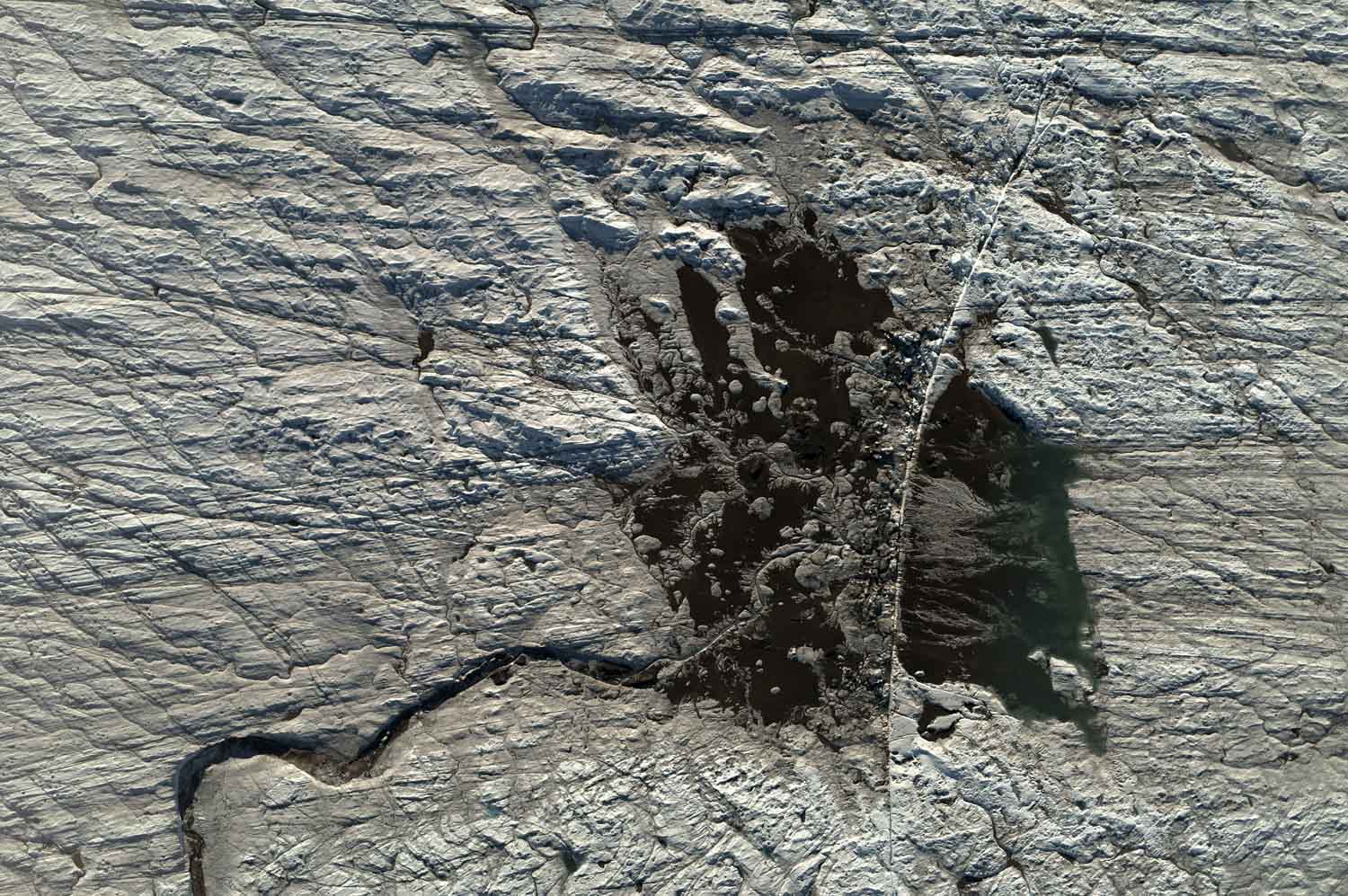

Arctic Aftermath (3rd Place, Jury Award)
Tilman Bucher, DLR & Angelika Humbert, AWI
This aerial image was acquired over the 79°N Glacier in NE-Greenland with the DLR Modular Aerial Camera System onboard the AWI Polar-6 aircraft on 20 August 2022 with a resolution of 12 cm per pixel. The image was captured to understand stress and time scales for the formation of ice-cracks and subsequent supraglacial lake drainage, and to validate models of the subglacial hydrological system by upscaling ground measurements to satellite observations. The drained lake resembling a fish is structured by meandering flow channels, remnant sediments coloring the body, the fins are depicted by finely branching structures. A bright sharp-edged fracture, visible as a 3m-step in the digital surface model, divides head and body; it drained the lake with the water now stored underneath the glacier. Fragments of ice in the vicinity of the crack were squeezed out during crack propagation. Cryoconite, a brownish material affecting the albedo of ice, is found along the margins of the structure.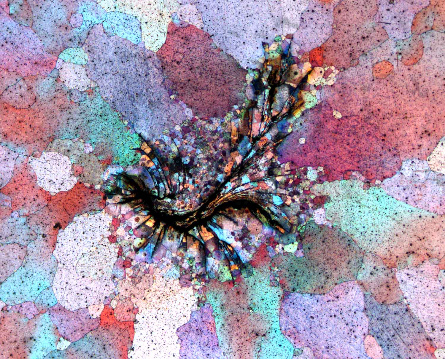

Magnesium Watercolour Whirl (1st Place, Participants' Choice Award)
Sarkis Gavras, HEREON
This image of a Magnesium alloy perfectly shows the amazing contrasts in grain size and orientation via a polarized light optical imaging technique within one sample. A naturally formed pore in the shape of a swirl is seen in this image. Countless miniscule grains of the order of 1 micron (10^-6 m) decorate this swirl shaped pore. The dramatic difference between the grain size of the grains shown forming at the pore and the larger grains further from the pore is a perfect example of how grain sizes can be modified via the presence of inclusions such as other particles or in this case the irregular morphology of a pore. Modifications of grain size and grain orientations are pivotal in designing tailor made materials which suit the usersʼ needs.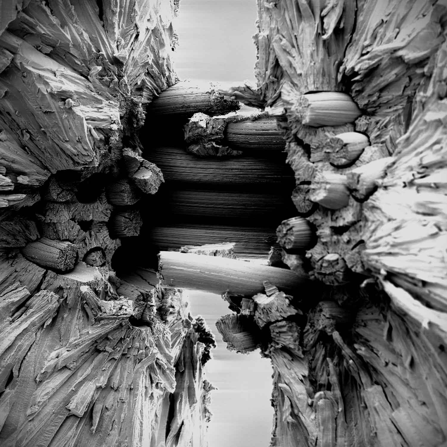

Fracture surface of yarn-based Wf/W (2nd Place, Participants' Choice Award)
Alexander Lau, FZ Jülich
Wf/W (Tungsten fiber-reinforced Tungsten) utilizes extrinsic toughening mechanisms to improve the mechanical properties of Tungsten, resulting in a pseudo-ductile behavior that expands its potential application range. The goal is to create a material that can withstand the high stress in potential future nuclear fusion reactors. The image shows the fracture surface of a new Tungsten fabrics made of yarns, which are stacked layer-by-layer into a solid composite using the chemical vapor deposition (CVD) of Tungsten hexafluoride and Hydrogen. The W-Yarns are infiltrated and encased in a CVD-W matrix.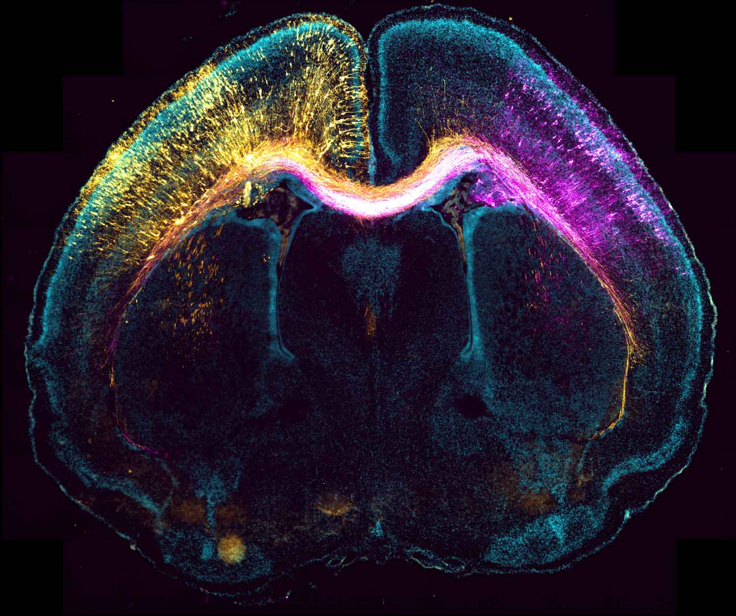

Stranger in the mirror (3rd Place, Participants' Choice Award)
Sebastian Dupraz (AG Bradke), DZNE
Neurons mirroring each other from the opposite hemispheres in the brain, they seemingly look the same but they differ in their very center. A pseudo-colored micrograph of a mouse postnatal brain previously subjected to bilateral in utero electroporation at an embryonic stage. The image was taken with a Plan-Apochromat 20X/0.95 N.A objective in an automated boxed LSM900 confocal microscope (Cell Discoverer 7; Zeiss) and pseudocolored in ImageJ. The right hemisphere shows control neurons (magenta), whilst in the left hemisphere (yellow), there are neurons expressing a peptide that inhibits microtubule nucleation from the centrosome. Bilateral in utero electroporation allows to follow the development of control neurons alongside modified ones and achieve an unambiguous evaluation of their behavior. With the help of images like this, scientists identified that microtubule nucleation from the centrosome coordinates the radial migration of cortical neurons but it is not involved in the formation or growth of their axons.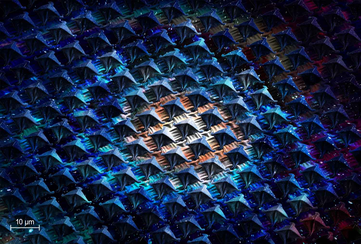

Small-pyramids Harvesting Big-universe Energy (1st Place, Public Choice Award)
Gan Huang & Bryce S. Richards, KIT
The universe is an abundant source of cold energy, and our image captures the potential of harnessing infinite cold energy to provide sustainable cooling for buildings, vehicles, solar panels, and more. To capture this energy, we’ve designed metamaterials with tiny micro-pyramids that absorb cold energy from the universe. Unlike the great pyramids in Egypt, these micro-pyramids are incredibly small, about one tenth the diameter of a hair. Our image showcases their intricate and beautiful design. It provides valuable information about their surface features, crucial for optimization. This image shows the importance of interdisciplinary research to meet clean energy demands. With this knowledge, we can develop efficient and sustainable cooling solutions for the future. The potential of cold energy harvesting with micro-pyramids is immense, and our research is just the beginning of a new era in sustainable energy.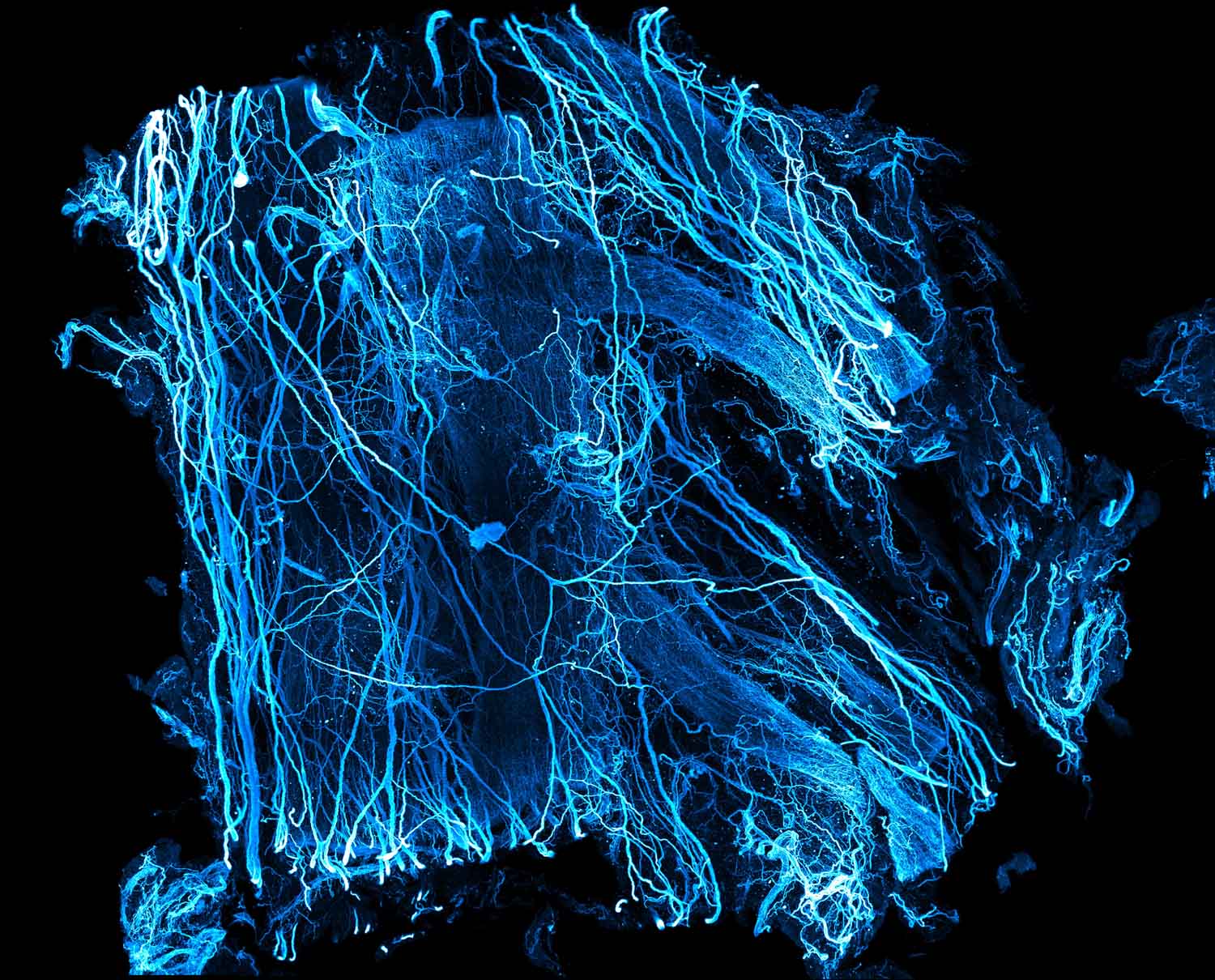

Innervation of human adipose tissue (2nd Place, Public Choice Award)
Hongcheng Mai, Jie Luo & Ali Ertürk, HMGU
Given the limited understanding of how sympathetic nerves innervate adipose tissue, we employed the deep tissue staining method. We used the Tyrosine Hydroxylase antibody as a marker to stain the sympathetic nerve fibers in centimeter-sized human adipose samples. After staining, we applied a tissue clearing technique, making the labeled adipose tissue optically transparent. This allowed us to use light sheet microscopy to obtain detailed single cell resolution images of the sympathetic nerve distribution within the human adipose tissue, enriching our comprehensive insight into the neuromodulation of adipose metabolism.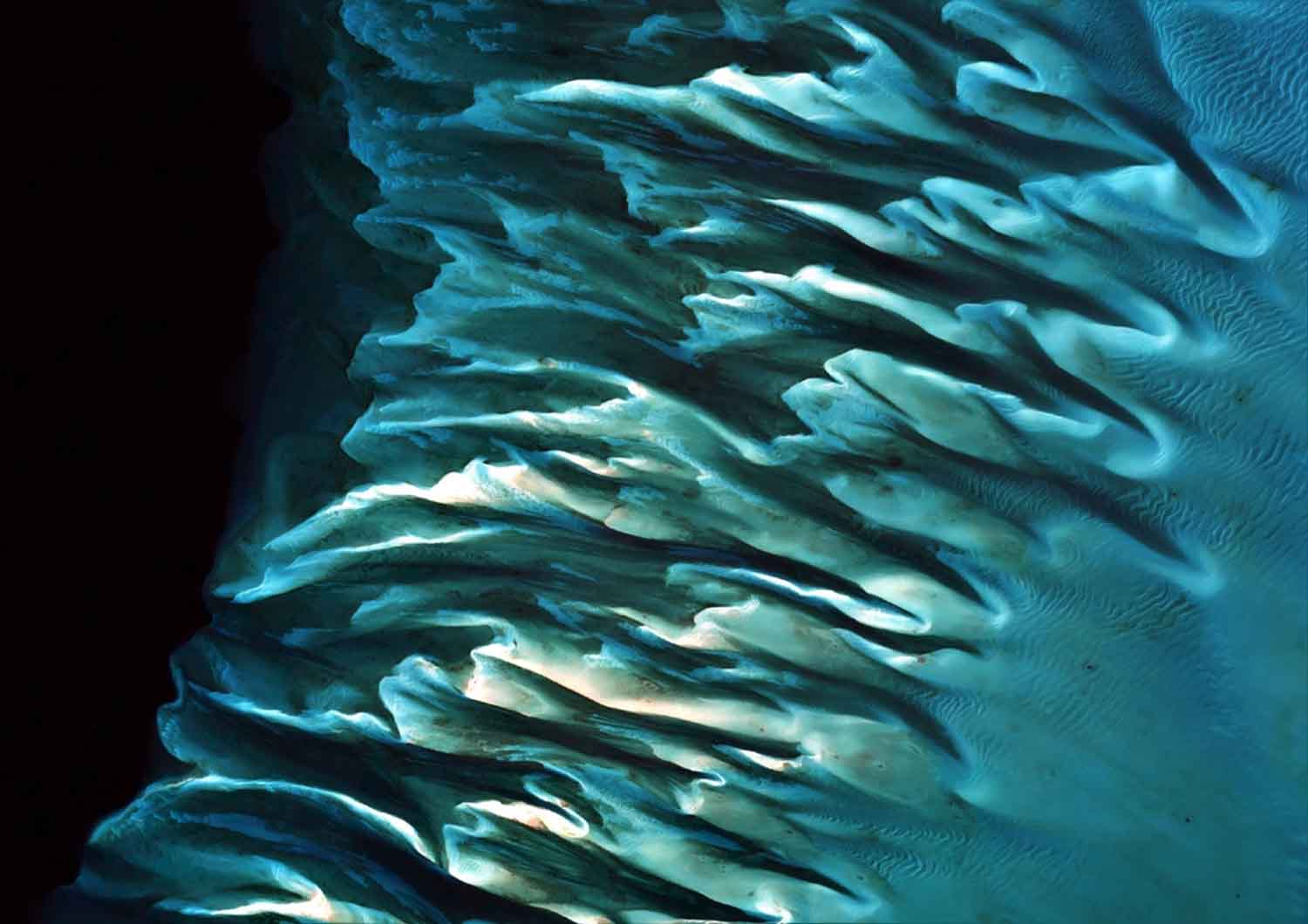

Glowing Bahamian seagrasses from space (3rd Place, Public Choice Award)
Alina Blume, Avi Putri Pertiwi, Chengfa Benjamin Lee, Spyros Christofilakos, Dimos Traganos, DLR & Global Seagrass Watch
Our submitted RGB image mosaic consists of 18,000 EU Sentinel-2 satellite images, acquired between 2017-2021 over the Great Bahamas Bank in the Bahamas. We utilized the Google Earth Engine platform for the mosaicking and ArcGIS 10.8 for the map. What makes our image so special is our multi-year mosaicking which allows an automated filtering of tropical optical obstacles such as dense clouds, turbidity and waves. This mosaic is part of a ~113,000 km² mosaic created to estimate the pan-Bahamian seagrass blue carbon. Our technology merges big satellite data analytics, machine learning, biophysical models, cloud computing with big field data — aiming to unlock the untapped potential of seagrasses for climate change mitigation. The Bahamas feature the world’s second largest national seagrass area, and had not been accounted for blue carbon prior to our efforts. Based on our estimates, Bahamian seagrasses could mitigate up to 68 times the country’s annual carbon emissions, rendering it carbon-neutral.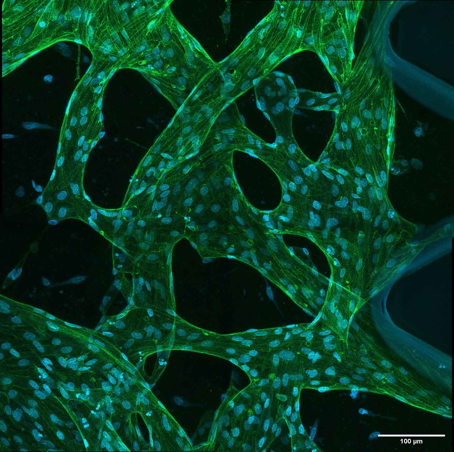

Blood vessels in a dish
Katharina Koch & Katja Meier, MDC
The image shows a vasculature which is built in a dish from single cells. Human cells from the umbilical vein were used. These basic building blocks of a vessel adhere to each other and form a tight vasculature through a protein called VE-Cadherin (stained in green). The nuclei of each cell is stained with Dapi. The vasculature forms in a 3D fibrin gel through self-assembly. With this in vitro method we can understand basic mechanisms of the process how blood vessels form and remodel, without using animals. It provides an alternative method for animal experiments. The 3D device was stained with fluorescent antibodies (VE-cadherin) and the image was captured with a Laser Scanning Microscope.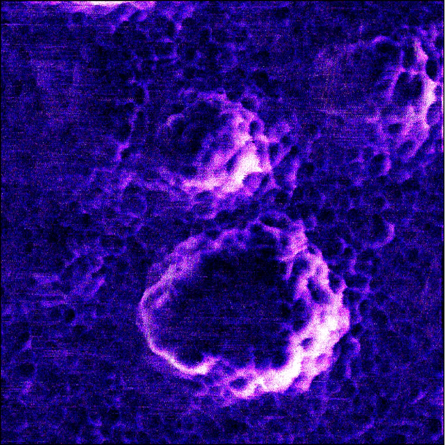

SARS-Cov-2 VLPs on TiO2 Photocatalyst
Heshmat Noei & Mona Kohantorabi, DESY
10.0 μL of SARS-CoV-2 stock containing 10.000 plaque forming units (PFU) (106 PFU/mL) was loaded on the center of single crystalline TiO2(101) so that the whole catalyst surface was covered. The sample was inactivated under UV light (wavelength was 256 nm, 10 cm distance to the light source) for 30 minutes. This sample was characterized by atomic force microscopy (AFM) technique. AFM imaging was performed using a CP-II instrument from Digital Instruments at DESY Nanolab. The AFM image was recorded under intermittent (tapping) mode, with a different scan size of 5.0 × 5.0 μm2 and 0.996 Hz scan rate. The SARS-CoV-2 VLPs adsorb on the titanium dioxide surface. The large particles are VLPs and the smaller features are the structural proteins denatured and adsorbed on the surface of TiO2 under UV irradiation. This is the first AFM image recorded from Sars-Cov-2 virus like particles on a solid photocatalyst surface.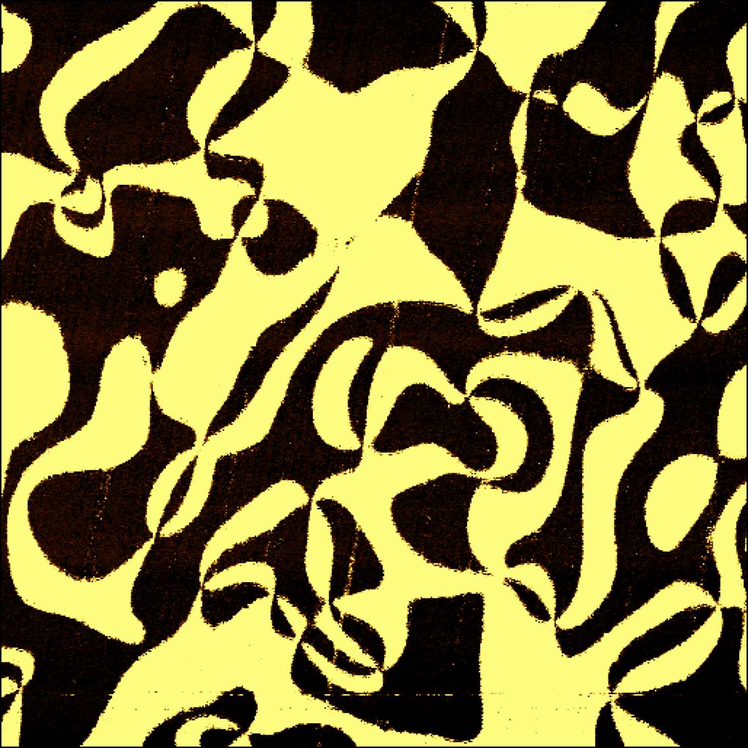

Ferroelectric swirl
Valentin Väinö Hevelke, Dong-Jik Kim & Catherine Dubourdieu, HZB
The image shows swirling ferroelectric domains of opposite polarization on the surface of a ErMnO3 single crystal imaged by piezo-response force microscopy (PFM). The image belongs to a study in which we have shown for the first time that ferroelectric domains can be visualized and characterized using a focused ion beam. In our study, we have used a focused beam of helium ions in the helium ion microscope (HIM). ErMnO3 was chosen as our test system because of its peculiar domain structure and its unusual domain wall configurations. The PFM image was taken in order to verify that the domains imaged by HIM were genuine. The open source software gwyddion was used for coloring the domains of opposite polarization.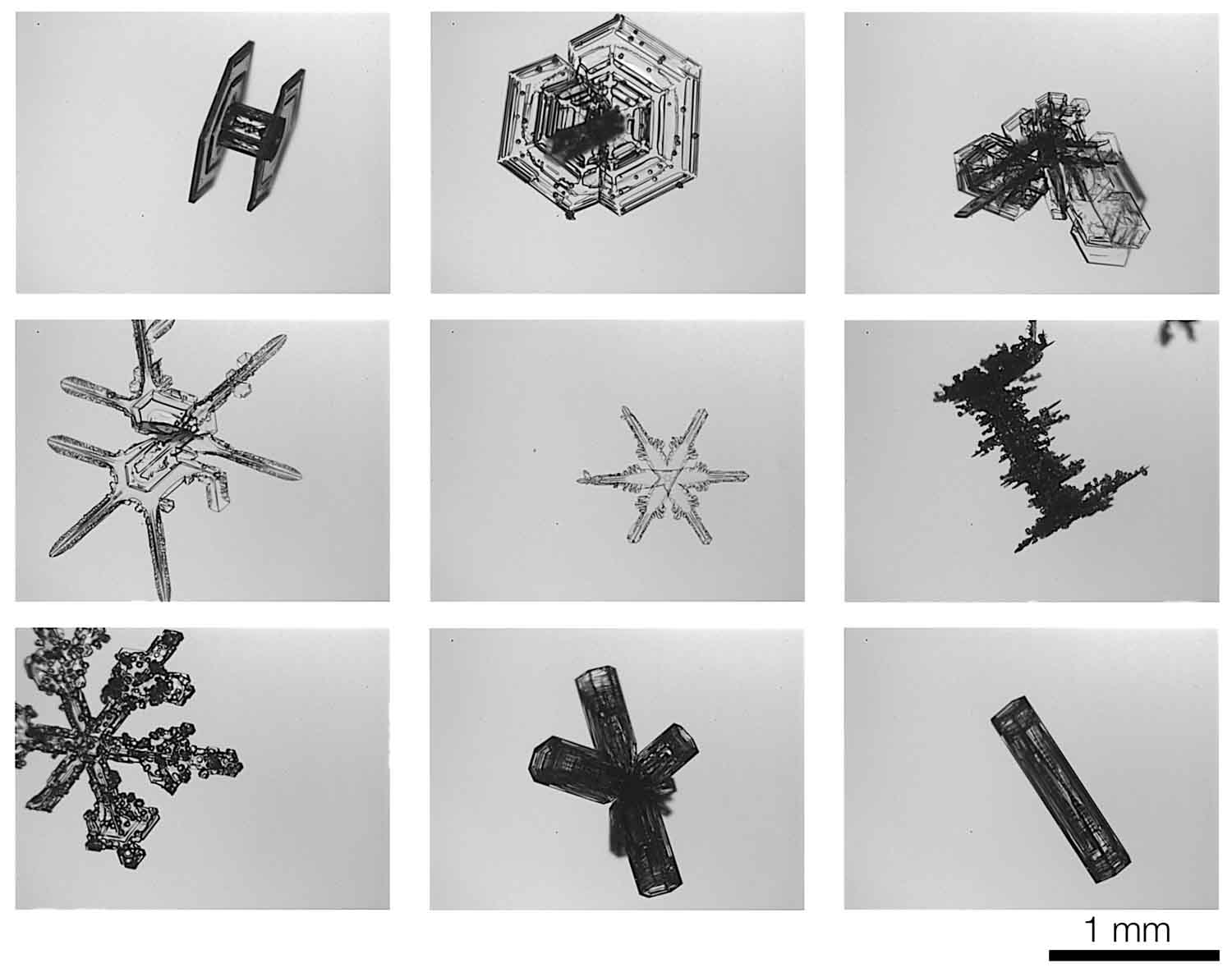

Secrets of Atmospheric Ice Crystals
Emma Järvinen & Martin Schnaiter, KIT
Ice crystals are breathtaking works of art crafted by nature. Their intricate designs and mesmerizing shapes adorn the world with dazzling beauty. However, beneath this splendor lies a hidden danger. Certain ice crystals have the power to trigger heavy snowfall, impacting transportation, commerce, and public safety. To better understand these phenomena and enhance snowfall forecasting, a team of international scientists embarked on a comprehensive study. Using advanced technology like the PHIPS airborne cloud instrument, they captured high-resolution stereoscopic images of single ice crystals. These images revealed the microphysical properties of snowstorm embryos, offering valuable insights to improve numerical weather models. By pinpointing the precise locations of these crucial embryos, lives can potentially be saved through enhanced forecasting capabilities. This submission showcases the delicate beauty of nature and underscores the importance of this research endeavor.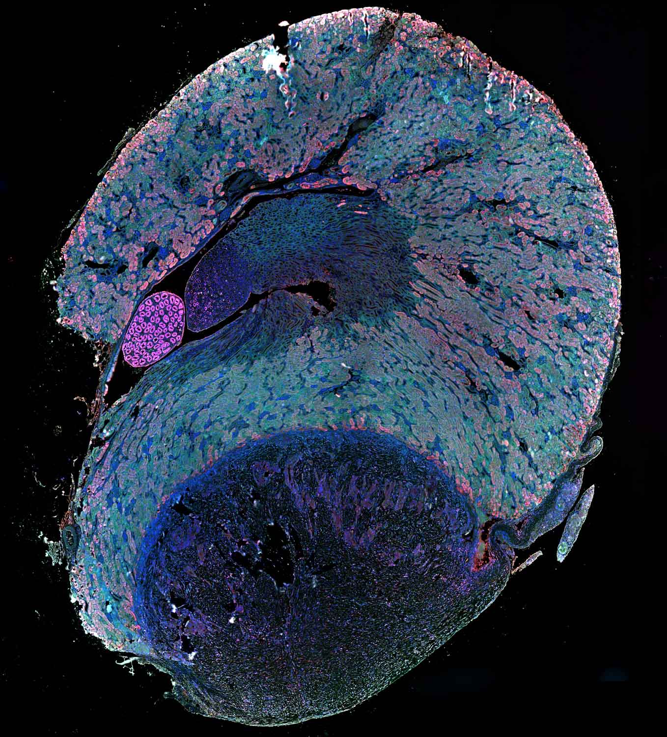

Embryo
Olawale Johnson Ogunsile, HGMU
Despite marked progress in clinical islet transplantation in restoring physiologic glucose homeostasis, one of the several obstacles precluding its widespread use in the treatment of type 1 diabetes is islet graft destruction and transplant failure. Islet death and rejection are mediated by T cells infiltrating the graft area after transplantation. This means that alloislets graft recipients must be placed on a long-term immunosuppression regimen to prevent the graft from getting rejected. However, using these immunosuppressive agents come with undesirable side effects including chronic toxicity, risk of infection, and malignancy. Our aim is to characterize the T cells invading the islets graft area post-transplant to understand their role in the rejection process. This knowledge can potentially lead to the development of future rejection prevention strategies that do not rely on long-term systemic immunosuppression.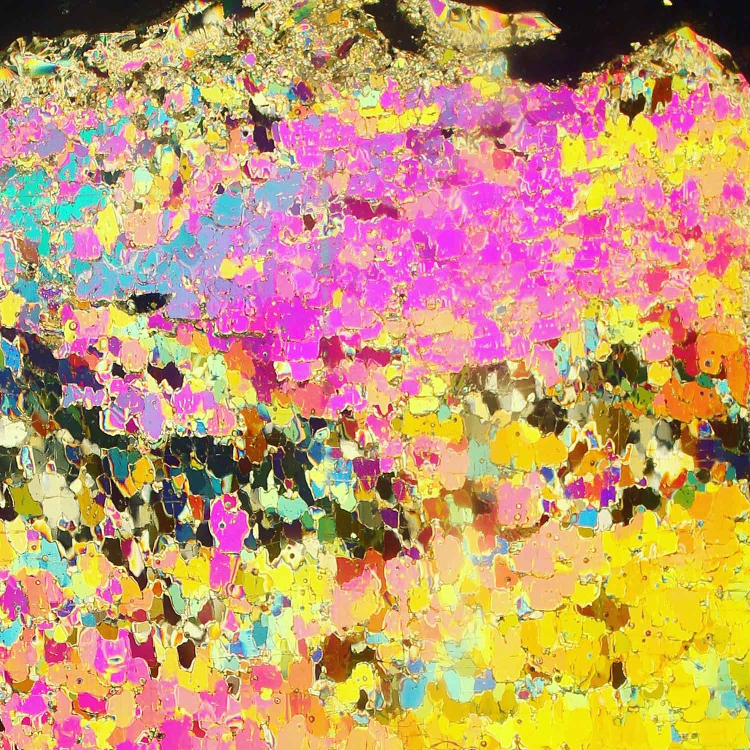

This is sea ice
Anja Rösel, DLR & Polona Itkin, UiT - The Arctic University of Norway
This photograph shows a longitudinal thin section of a sea ice core. It has the size of approximal 8 cm x 8 cm. This thin section is made out of a 7-8 mm thick slice of an ice core form the Arctic sea ice, glued with water on a glass plate and processed by a microtome to a very thin layer of < 1 mm. To make the crystal structure of the individual sea ice crystals visible, the glass plate is illuminated with polarized light. Here we see a typical pattern for the so called granular ice. That means the ice has grown under “rough”, maybe windy, conditions. The knowledge of the crystal structure of the sea ice is important to better understand and to model the light transition through the sea ice into the ocean, which is, for example, of relevance for the phytoplankton growth in the Arctic Ocean.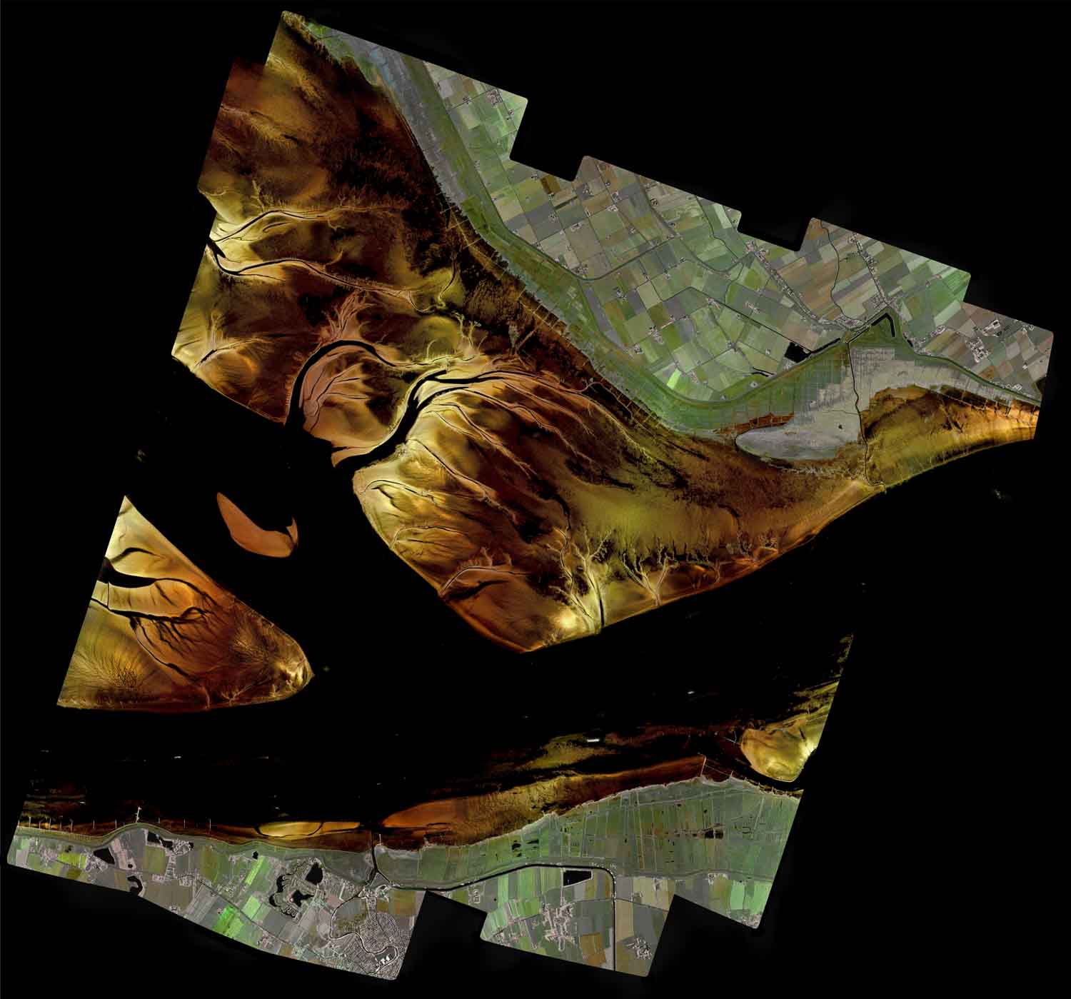

Medem Rinne seen with Radar Eyes
Jens Fischer, Joel Amao-Oliva, Muriel Pinheiro, Marc Jaeger, Rolf Scheiber, Martin Keller, Ralf Horn & Andreas Reigber, DLR
Acquisition: This image is a mosaic of high-resolution radar images taken by DLR’s airborne Synthetic Aperture Radar (SAR) system F-SAR over the Elbe estuary near Hamburg, Germany. The extent of the scene is 14 km x 12 km at a resolution of 50 cm x 50 cm per pixel, and the total acquisition time was about 2 hours. What makes it special: In this image, we combine Synthetic Aperture Radar (SAR) and High Dynamic Range (HDR) processing for the first time. We decoupled land and tidal flats so that both parts of the image could be optimized independently. Colors: The image was taken at a wavelength (9 cm, S-band) where humans cannot see. We therefore obtain gloss effects on structures (sandbars and meadows) that would not shine with human vision. The colors of the image correspond to the polarimetric properties of electromagnetic waves and they are chosen so that house roofs appear red, vegetation green and sand in golden colors.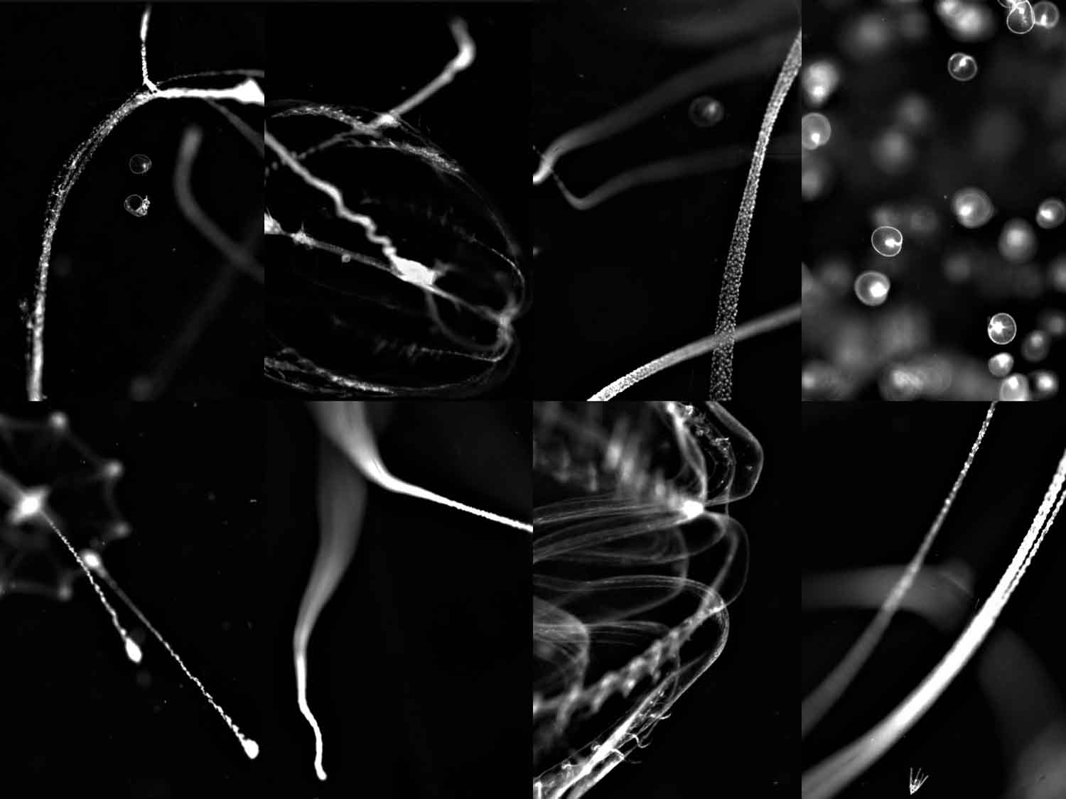

Looking for Noctiluca
Katharina Kordubel, HEREON
These images represent waters dominated by Noctiluca scintillans (top left) and comb jellies, and were captured by the CPICS (Continuous Plankton Imaging and Classification Sensor), deployed in summer 2022 in Helgoland to sample N. scintillans blooms, which have globally spread, become more intense and frequent over the last decades. This is problematic, as N. scintillans is a trophic dead-end and food chain disruptor, causing ammonium toxicity and oxygen depletion. Due to rapidly developing and collapsing blooms and limitations in traditional techniques (nets, bottles), the knowledge about bloom dynamics and associated impacts is scarce. For the first time, an entire N. scintillans bloom was recorded. This revealed feeding, reproductive and interactive behavior in-situ, and validated laboratory-based knowledge. These data will improve our understanding about biology, functional role and dynamics under future conditions of this important organism.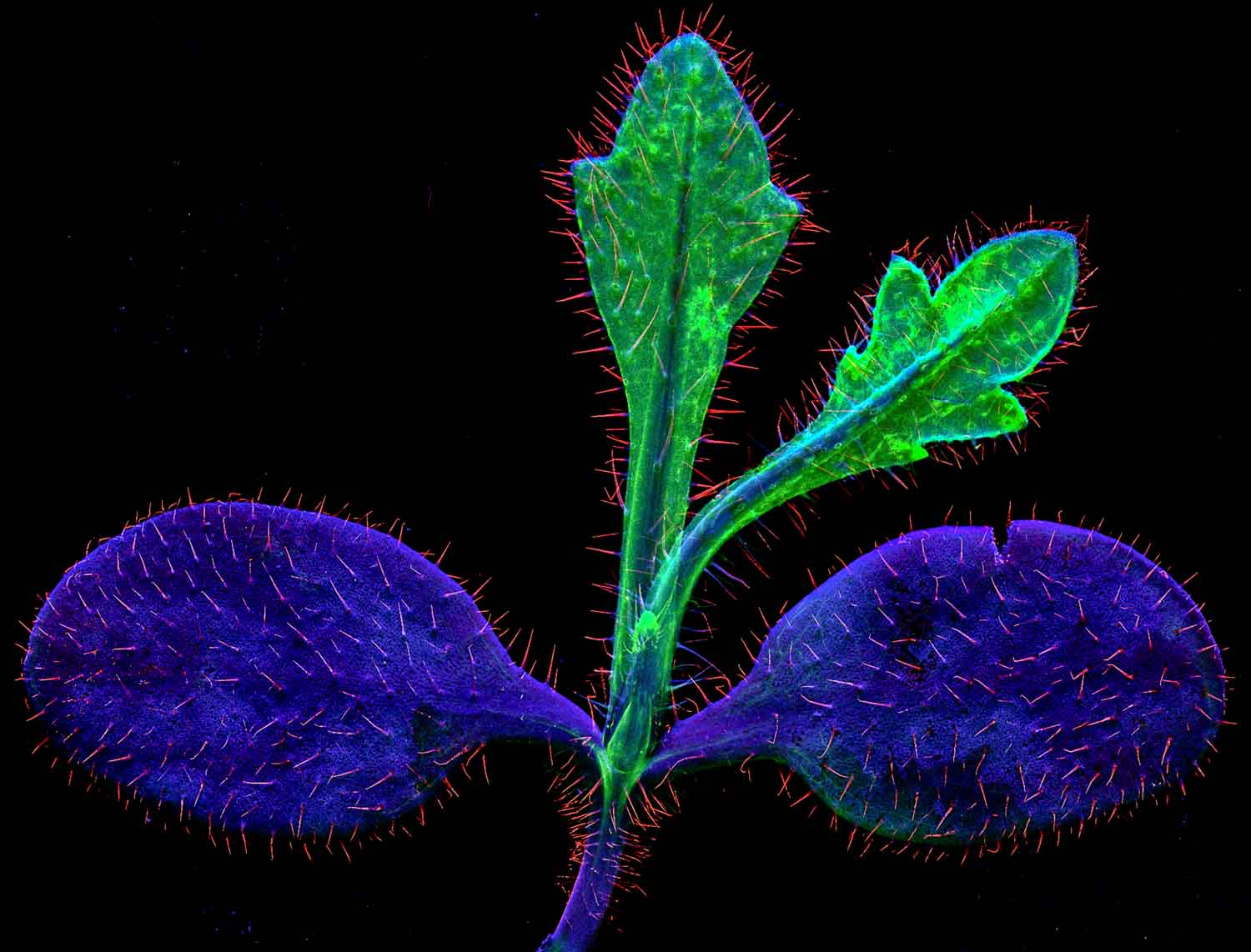

The deadliest plant?
Kathryn Spiers, Dennis Brückner, DESY; Antony van der Ent; Wageningen University & Research, The Netherlands & Mirko Salinitro, University of Bologna
This plant of the same family (Brassicaceae) as cabbages and cauliflower accumulates thallium in its leaves. Biscutella laevigata grows on the mine spoils of the ancient mines near St Laurent le Minier in Southern France. It can contain over 30,000 mg/kg thallium in its leaves. This image is a synchrotron micro-fluorescence elemental map acquired at beamline P06 of DESY and shows calcium in red, thallium in green and potassium in blue color. This B. laevigata seedling has toxically-high levels of thallium in the two youngest leaves, while the lower levels of thallium in the older, rounder leaves allows potassium to dominate the signal. The leaves are covered with calcium-rich hair-like trichomes. Thallium contamination of the environment is common around former zinc-lead mines and smelters, and this plant can be used in an innovative method for decontamination called ‘phyto-extraction’ to remove thallium from the soil.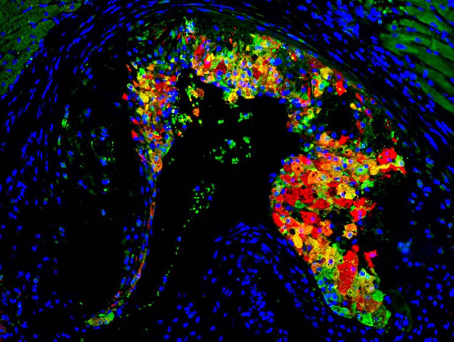

Colorful killer
Fatimunnisa Qadri & Bernadette Nickl, MDC
Shown is an atherosclerotic plaque formed in the valves of a mouse heart during a high-cholesterol diet. The plaque is deposited over the smooth muscle cells. Cholesterol from blood accumulates on the valves and is taken up by macrophages. Cholesterol-filled macrophages form a plaque which narrows the vessel lumen. Rupture of such plaques may cause myocardial infarction and stroke. We tested whether the protein Gpnmb is involved in cholesterol-induced plaque formation. A mouse heart section was stained using antibodies against a macrophage cell surface marker, CD68 (green), and a second antibody directed to Gpnmb (red) and imaged under a fluorescence microscope (Keyence BZ-9000). Blue dots represent cell nuclei. Interestingly, Gpnmb is seen in macrophages (yellow) and also other cells (red), implying Gpnmb may have a role in atherosclerotic plaque formation.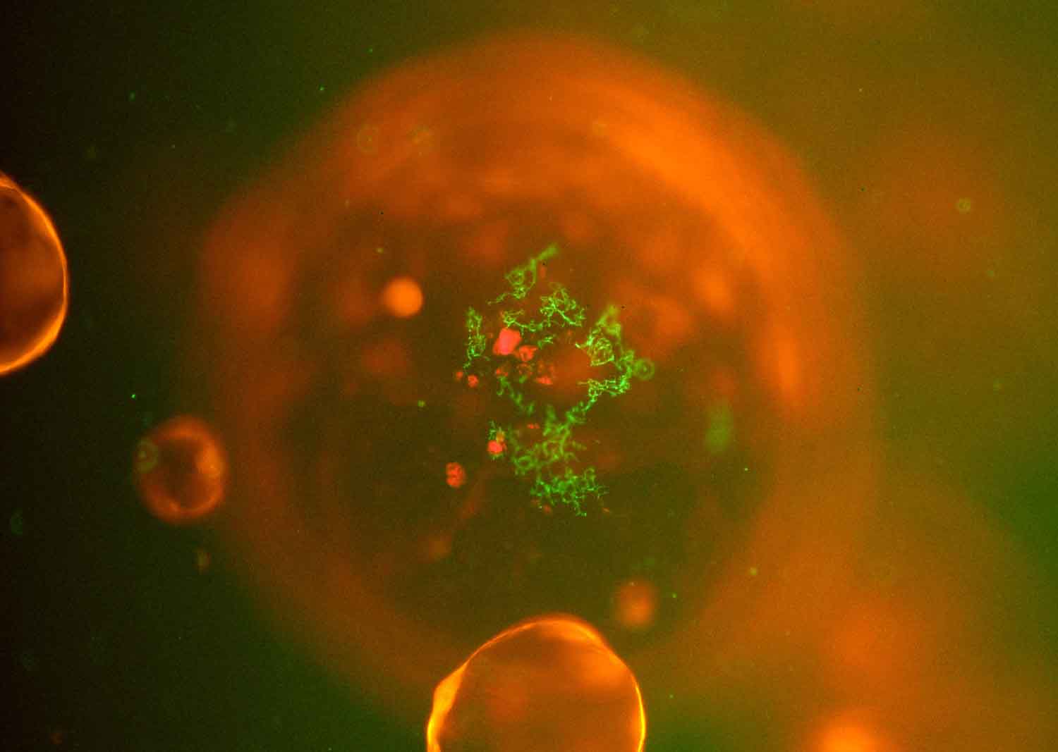

Understanding host-microbe interactions using patient-derived organoids microinjected with bacteria
Lena Schorr, DKFZ
Reporter bacteria were microinjected into the lumen of lentiviral labeled organoids derived from the colon. The image was taken with a fluorescence microscope and is therefore unique, since such a microinjection setup is rare to study bacteria-host interactions and ultimately enables to gain more functional insights into host-bacteria interactions.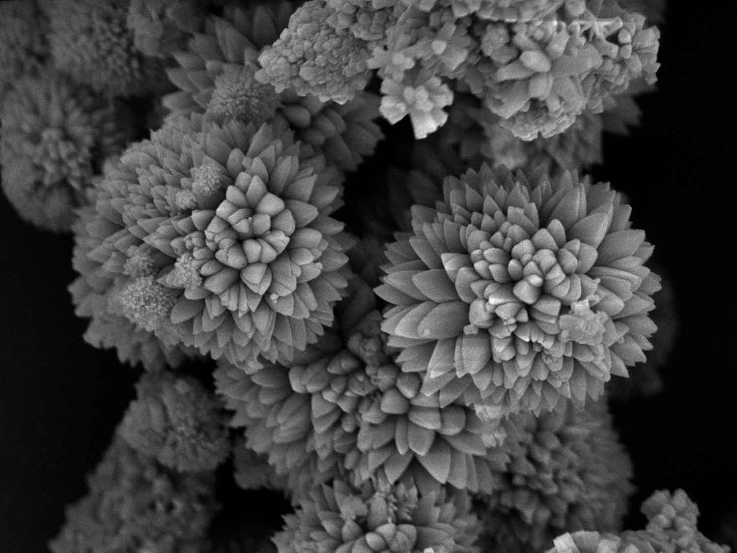

Rhodium Chrysanthemum
Martin Etter, DESY
An organic-inorganic Rhodium salt whose crystals grows in the shape of Chrysanthemum flowers. The image was taken by a scanning electron microscope and the colors are unchanged. The size of the right bigger flower has an approximate diameter of 10 micrometers. At the time when the image was taken, the underlying crystal structure of this Rhodium salt was unknown. And from the outer appearance of the crystal shape it was hoped that some information about the atomic crystal structure symmetry could be retrieved. And indeed, the shape of a single leaf suggested a crystal system with angles different from 90 degrees. In the end, the crystal structure could be solved by X-ray diffraction methods with a rhombohedral symmetry. Currently the material is still investigated regarding its catalytic properties.