Best Scientific Image Contest 2024
Once again, we invited the Helmholtz community to showcase their exceptional imaging capabilities. This year, we received 127 high-quality images. View the top selections in our virtual gallery where the best 20 images of 2024 are featured!
Gallery
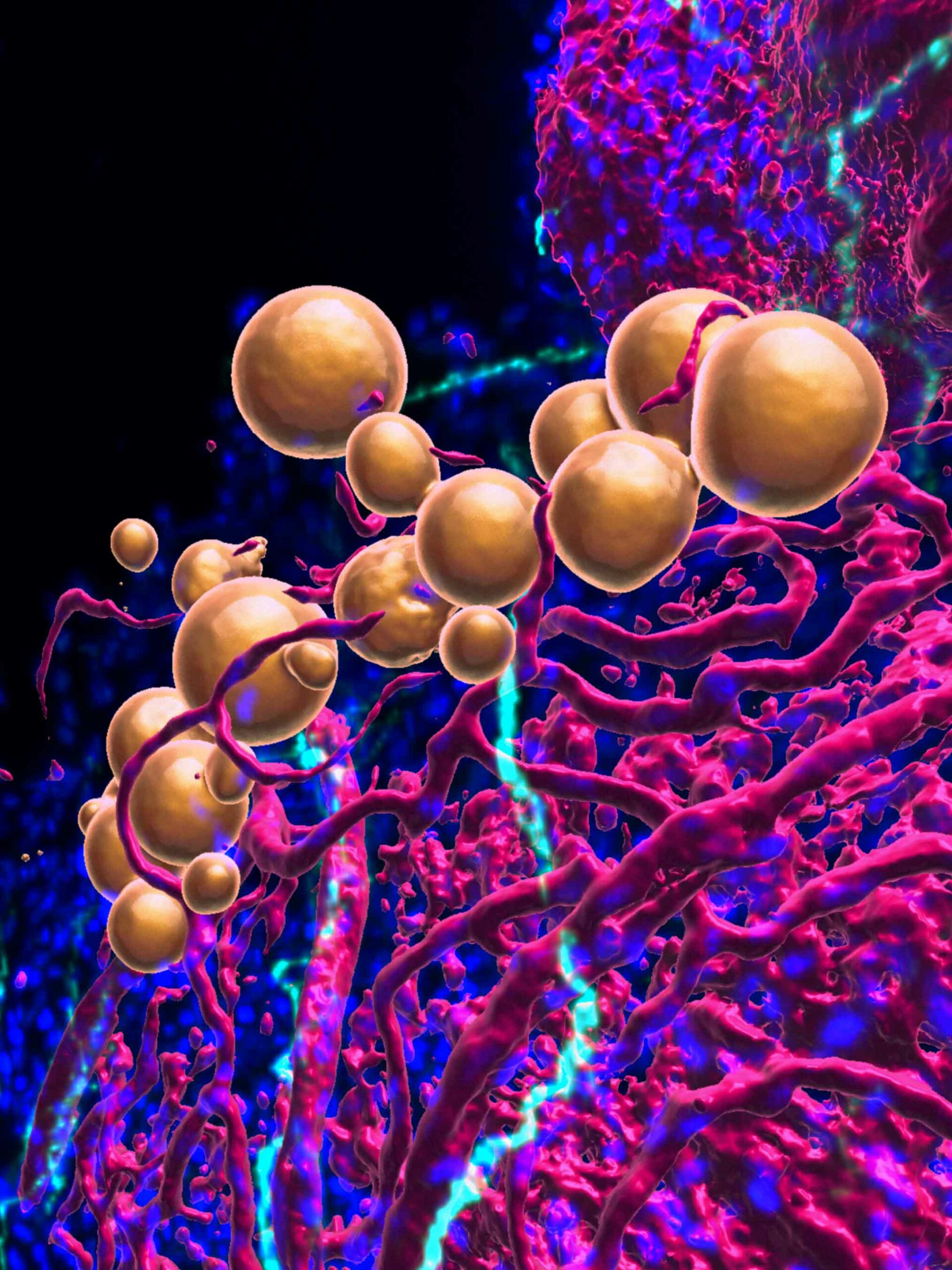

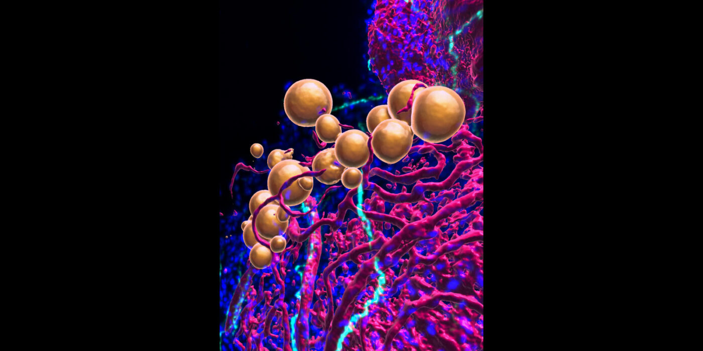
More than just fat (1st place Jury Award)
Paul A. Morocho Jaramillo (AG Sawamiphak), MDC
This confocal image explores the epicardial adipose tissue (eAT) in an adult zebrafish heart, a subject of study that has been less explored due to the scarcity of suitable animal models. Zebrafish eAT closely resembles the metabolically active fat found in humans, being highly innervated and vascularized by coronary vessels. This work is particularly notable for identifying a cardiac fat depot in zebrafish that mirrors the localization and architecture of human beige eAT, achieved through tissue-clearing and precise staining methods to reveal optically transparent adipose tissue. The significance of this image lies in its demonstration of the value of using model organisms, such as zebrafish, to gain insights into adipocyte biology and its implications for human health. The detailing in the image is brought out through staining: BODIPY for lipids/adipocytes in yellow, DAPI for nuclei in blue, a transgenic line kdrl:HRAS-mCherry for the coronary vascular system in magenta, and acetylated tubulin for innervation in cyan.

Decontaminating metal pollution with hyperaccumulators (2nd place Jury Award)
Kathryn Spiers, Dennis Brückner, DESY, Julien Jacquet, Gabrielle Michaudel, Econick SAS (France) & Antony van der Ent, WUR (The Netherlands)
This image was produced by merging three images, each on a different color channel, showcasing chemical elements or absorption that were obtained through Synchrotron X-ray Fluorescence Microscopy at the P06 beamline, PETRA III, DESY. This technique was applied to study hyperaccumulators, rare plants adept at concentrating metals in their cells and tissues, to deepen our understanding of the mechanisms behind metal hyperaccumulation. Remarkably, this experiment produced the first high-resolution elemental maps from fresh, hydrated flowers of such a plant. These maps are crucial for uncovering the cellular localization of hyperaccumulated elements, offering insights into their transport and storage mechanisms within the plant.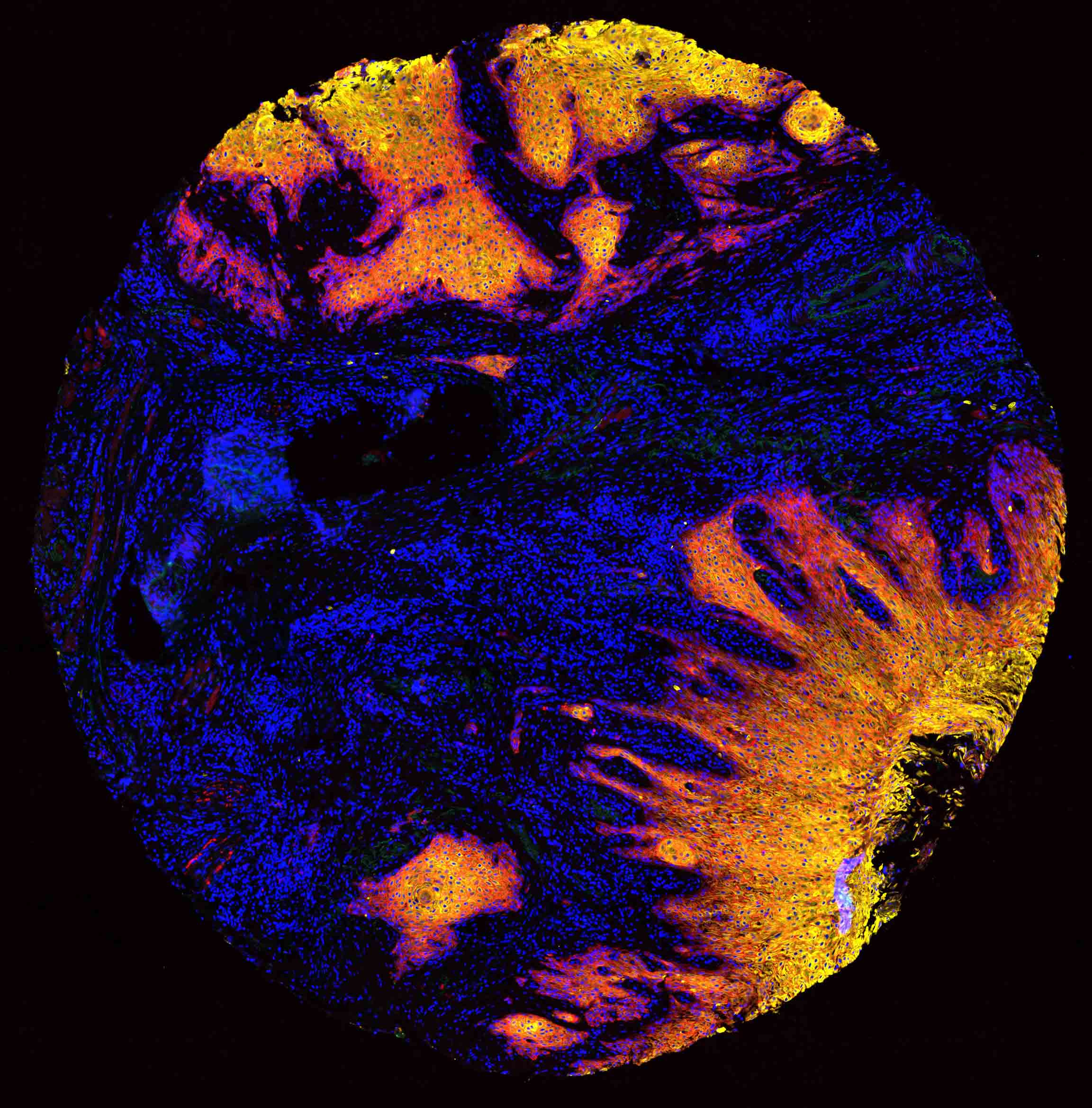

Conquering the cancer inferno (3rd place Jury Award)
Sonja Fritzsche, Simon Schallenberg, José Nimo, Fabian Coscia, MDC
Captured using an Axioscan 7 Slidescanner, the image showcases tissue stained with AlexaTM fluorophore conjugated primary antibodies, a technique to study treatment resistance in head and neck cancer. What sets this image apart is the vivid depiction of cancer cells invading healthy tissue, like an inferno. Through such detailed visualization by means of immunofluorescence microscopy, the goal is to unravel how cancer cells interact with the surrounding microenvironment, contributing to treatment resistance.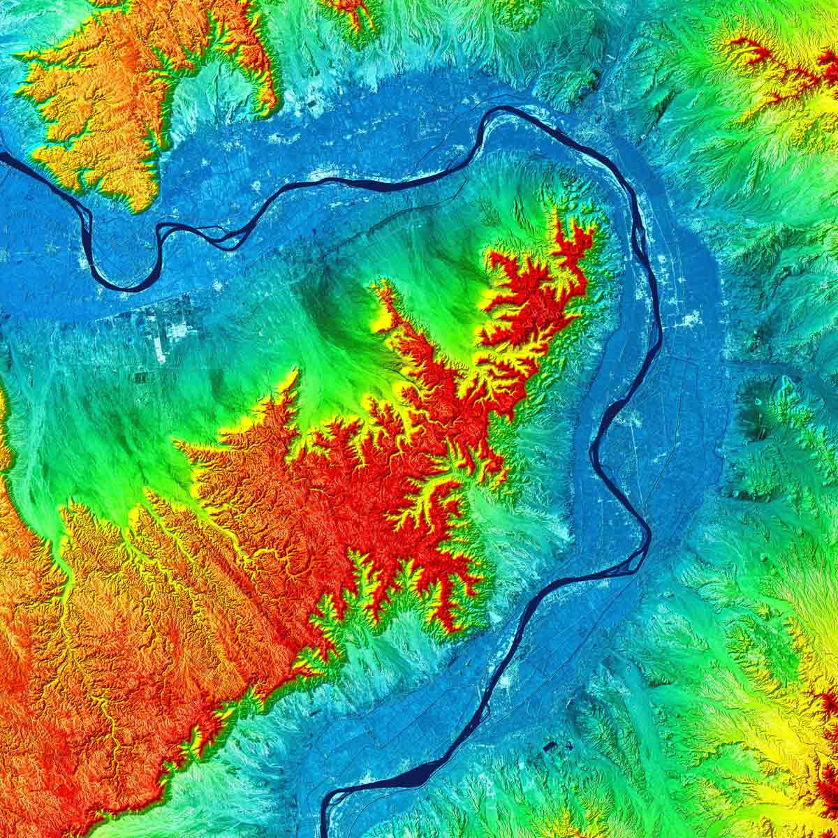

The kings valley tanDEM-X DEM
Markus Bachmann, DLR Oberpfaffenhofen
The data for this Digital Elevation Model (DEM) was captured using the TanDEM-X Synthetic Aperture Radar (SAR) interferometer, processed into DEM data with water bodies smoothed out for a flat appearance. This processed data was then visualized as a topographic map, with special attention to flat terrains where height information and SAR amplitude data were merged to reveal details like urban structures. This image was particularly focused on the Nile river valley, shown in dark blue, to assess the accuracy of water body extents crucial for hydrological modeling. What makes this image extraordinary is the careful adjustment of the color bar and scaling, offering a scenic view of the Earth's topography and making it easier to understand the landscape intuitively. This imagery is vital for hydrological applications, which depend on accurate representations of water bodies and river flow directions. The accuracy of the TanDEM-X DEM and the process of water body editing were evaluated through this image. Moreover, this image, showcasing the Valley of the Kings in Egypt, was highlighted during the 100th anniversary of the opening of Tutankhamun's tomb, as part of the TerraSAR-X/TanDEM-X Highlight Images, celebrating its significance in both scientific and historical contexts.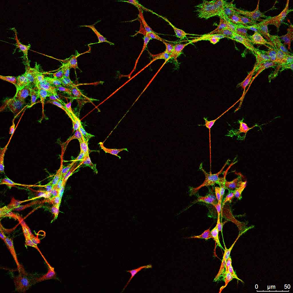

Friendship between brain tumor cells
Julia Sundheimer, DKFZ, KITZ & Kendra Maaß, Kristian Pajtler, DKFZ, KITZ, UKHD
This immunofluorescence image showcases pediatric brain tumor cells (ependymoma), vividly colored with DAPI (blue) for nuclei, actin (green) and tubulin (red) for the cell's cytoskeleton. The purpose of the image was to delve into the characteristics of the connections between these cells, revealing an intriguing aspect: no distance is too far for these cells to connect, mirroring the resilience of a strong friendship that endures despite physical separation. This image is particularly special as it beautifully illustrates how cells maintain connections even when spaced far apart, offering a visual metaphor for enduring connections and providing insights into cellular behavior and interaction.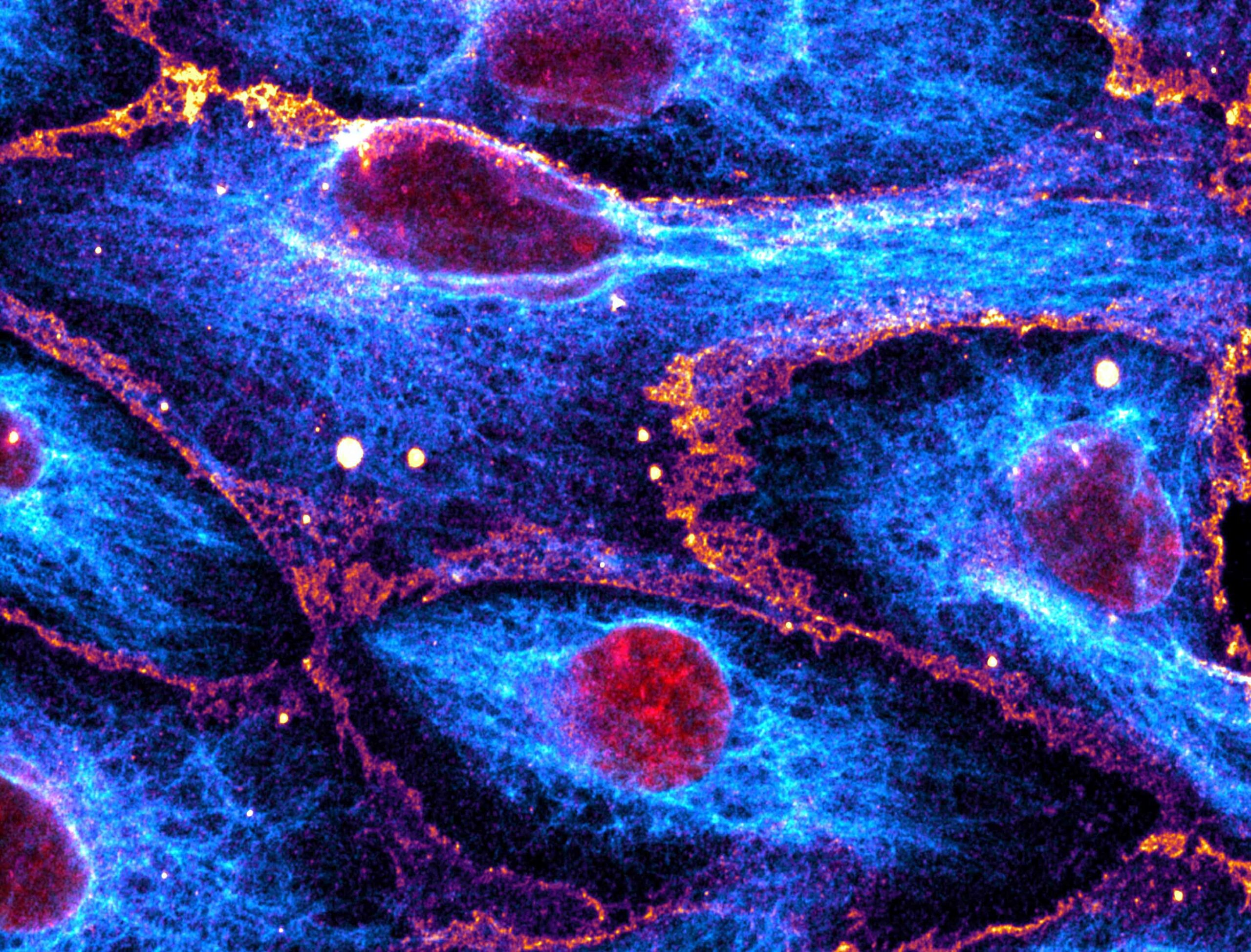

Watched over by vimentin's embrace
Emir Bora Akmeric & Julia Kraxner, MDC
Utilizing immunofluorescence staining and confocal microscopy, the image was captured to reveal the dynamics of vimentin fibers in endothelial cell monolayers under simulated blood flow conditions, seeded on Ibidi perfusion slides. Color Lookup Tables (LUTs) were applied: Red for DAPI (DNA), MPI-Inferno for VE-Cadherin antibody, and Cyan Hot for Vimentin antibody immunofluorescence, to distinguish the different elements clearly. This image is remarkable for displaying the vimentin networks' morphological shift, aligning in the direction of the simulated flow, a phenomenon along with its nuclear "blanketing" effect that had not been observed before. The visualization of flow-induced fiber alignment suggests a protective mechanism for cell nuclei, highlighting potential connections between cytoskeletal biophysics, epigenetics, and vascular health. Captured with a Zeiss LSM 980 confocal microscope using a 63x 1.4NA oil objective in SR-4Y multiplex airy scan mode for super-resolution imaging, three separate excitation/emission filters and laser configurations were utilized to image different molecular targets. These were then overlaid using differently colored lookup tables. A total of 32 z-slices, 150 nanometers apart, were merged into a single image through Z-Projection, offering an in-depth view into cellular responses to mechanical stimuli.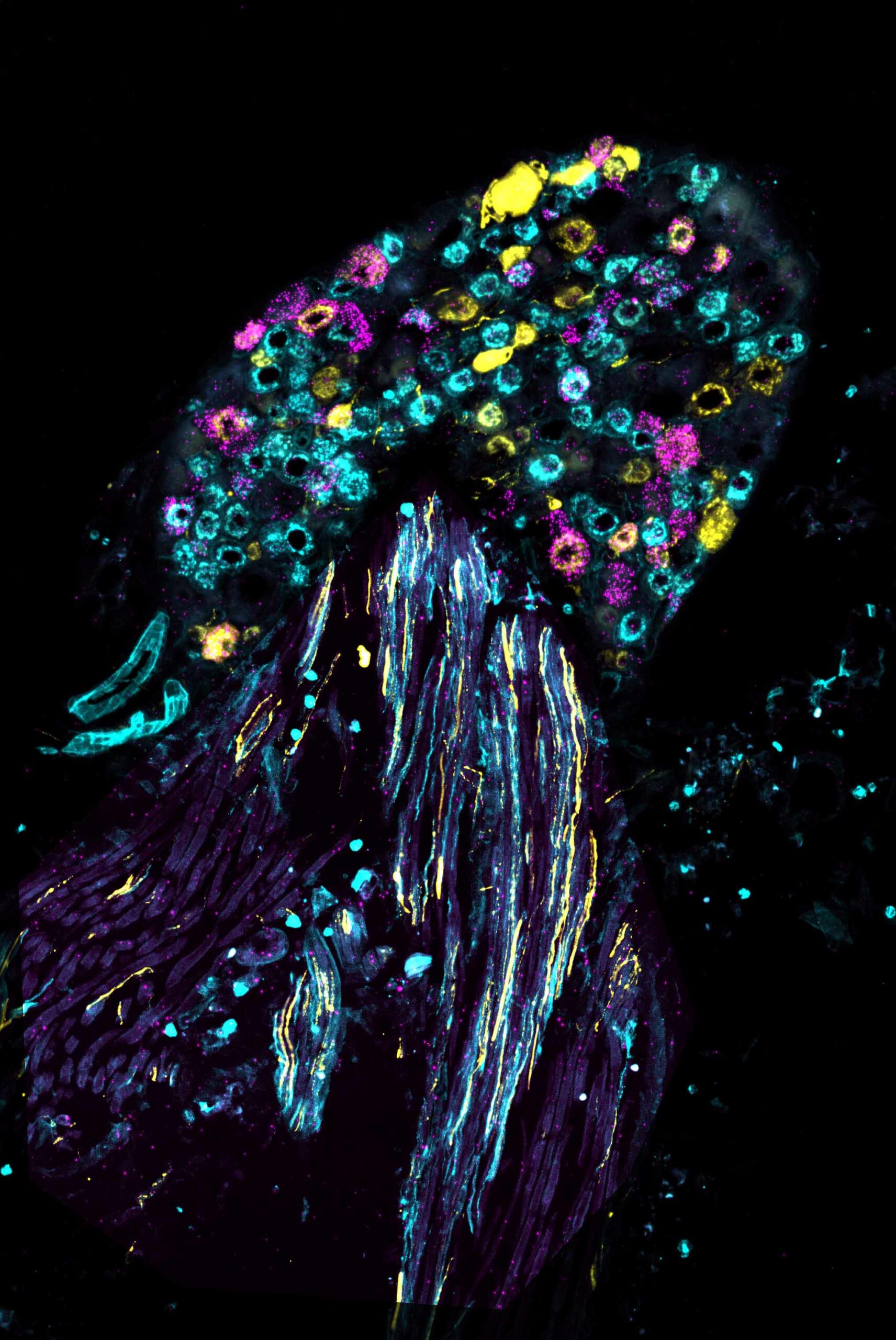

Pain neurons or jellyfish? (1st place Public Choice Award, 2nd place Participants' Choice Award)
Athanasios Balomenos, Lin Wang, MDC
Captured using an Olympus IX83 and CSU-W1 spinning disk confocal system, this image was “colored” with fluorophores through a combination of RNAScope and fluorescent immunohistochemistry to label RNA and protein molecules. It showcases a mouse dorsal root ganglion (DRG) highlighting different populations of sensory neurons and visualizes the overexpression of the Stomatin-like protein-3 (Stoml3) in magenta within pain neuron subpopulations marked in yellow and cyan. What sets this image apart is its striking resemblance to a jellyfish swimming in the sea; the main body of the DRG, with sensory neuron somata, mirrors the jellyfish's bell, while the fibers resemble its tentacles and oral arms. This visualization elucidates the specific overexpression of the Stoml3 molecule in pain sensory neurons, implicating its significant role in neuropathic pain in mice. The discovery that treatment with Stoml3 inhibitors can rescue this phenotype highlights a potential therapeutic target.

Star clinopyroxene glomerocrysts
Louis-Maxime Gautreau, GEOMAR
Captured through a petrographic microscope using transmitted light, this image showcases the unique characteristics of magmatic rocks from Conical Seamount, a submarine volcano located in north-eastern Papua New Guinea. What makes this image particularly captivating is the almost perfect star shape observed within the rocks, sparking widespread interest and sparking conversations beyond the scientific community. This highlights how a single, simple image of specific crystal formations can significantly enhance scientific outreach to the general public. From such images, scientists aim to glean insights into the processes occurring within the magma chamber beneath the submarine volcano. Due to the image's large size, a specialized software linked to the microscope was employed to capture multiple images at regular intervals. These images were then meticulously juxtaposed to create the final comprehensive image.

Gut ring (1st place Participants' Award)
Daniel Postrach, DKFZ
Using confocal fluorescent microscopy, this image of a fixed mouse intestine was captured, highlighting the organ's detailed structure through immunostaining techniques that illuminate the cell membrane, blood vessels, and actin fibers. This image was taken as part of a study focusing on the intricate sub-anatomical architecture of the intestine. What sets this image apart is the clear depiction of a complete ring of the intestine, showcasing all its villi and structures and revealing the organ's fascinating complexity. Through such detailed imaging, researchers aim to gain a deeper understanding of the cellular organization within the intestine, with a particular interest in the layout of the blood vessels.

A comb for quantum states
Sven Velten, Lars Bocklage, Ilya Sergeev, DESY & Ralf Röhlsberger, HI Jena
Recorded at beamline P01 at PETRA III, the interference pattern displayed is a vibrant representation of single photon counts, visualized on a logarithmic scale from dark to bright colors, showcasing variations in time and energy. This imagery was captured to illustrate the functioning of a frequency comb, which consists of evenly spaced quantum states, playing a crucial role in the storage and retrieval of single photons — the quantum particles of light. What sets this image apart is its depiction of a frequency comb measurement at X-ray energies for the first time, highlighting its potential as a quantum memory capable of storing X-ray photons. This interference pattern provides detailed insights into the properties of the quantum states held within the comb, offering a histogram of single photon decay events mapped across arrival time (vertical axis) and the spacing of the quantum states (horizontal axis).

Cells in dialog
Kamil Lisek, Svea Beier, Ilan Theurillat, Tancredi Pentimalli & Nikolaus Rajewsky, MDC
Using specialized techniques to label proteins, this photo captures cancer associated fibroblast (CAFs) cells in red and tumor cells in cyan and green, employing specific antibodies for color differentiation. The purpose behind this image was to study the reciprocal interactions between tumor cells and their microenvironment. What makes this image particularly remarkable is the captured cell interactions and their distinctive shapes, highlighting a unique moment in cellular communication. Through the analysis of such images, the goal is to understand the mechanisms of cell communication more deeply. This image was created using a Leica DMi8 Thunder imaging system, illustrating the power of advanced microscopy in revealing intricate details of cellular behavior and interaction.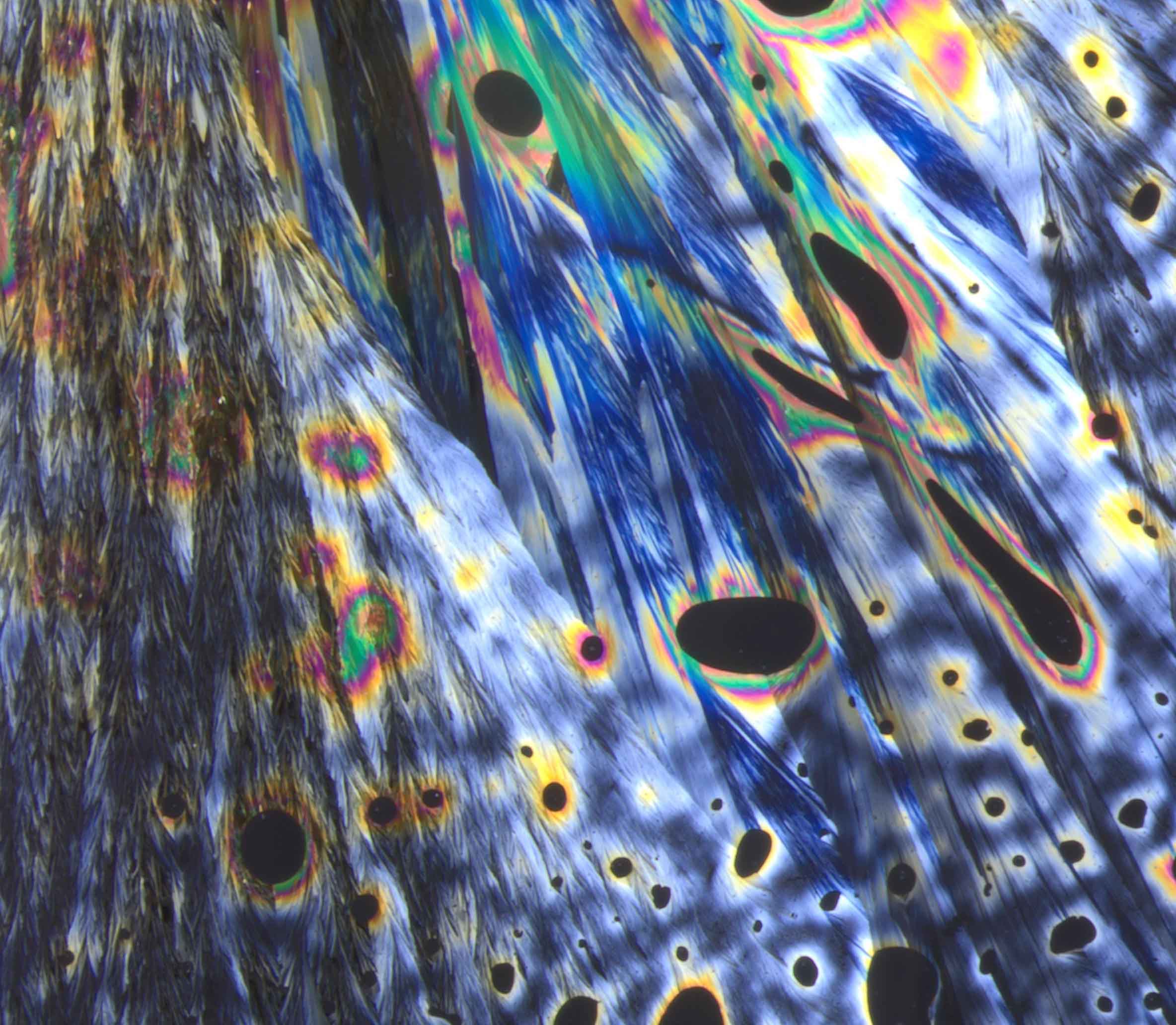

Azobenzene peacock feather crystals (3rd place Public Choice Award)
Sreevidya Thekku Veedu, Patrick YA Reinke, DESY
Captured through an Olympus SZX16 light microscope equipped with a polarization filter, this image showcases the striking microscopic view of thin multi-crystalline layers of azobenzene, a molecular photo switch capable of reversible isomerization between its trans and cis forms upon exposure to light stimuli. This characteristic, fundamental to azobenzene, renders it a promising candidate for applications in photobiology and photopharmacology due to its photo switchable behavior. The image is particularly special because it employs a polarization filter to bring out vivid colors in the microscopic crystals, making them resemble peacock feathers. This visual effect is achieved without further editing, highlighting the natural beauty of the small organic compound crystals at magnification and completing an optical illusion with bright, peacock-like colors. Such ultrathin single crystals are being explored for their potential for time-resolved diffraction experiments with relativistic MeV electrons at DESY's ultrafast electron diffraction facility REGAE.

Human retinal organoid
Natalie Dumler, DZNE
Captured using the ApoTome 2 fluorescence microscope (Zeiss) at 10x magnification, this image shows light-sensitive photoreceptors in green and Müller glial processes in magenta within a human retinal organoid. This visual was part of an analysis aimed at investigating pathological mechanisms. The image is notable for featuring an organoid that, at 400 days old, exhibits no signs of neurodegeneration, which is remarkable considering that organoids typically mature in 200 days but may later undergo spontaneous degeneration. This longevity indicates the effectiveness of the organoid protocol in generating stable organoids for extended periods. From this image, two key scientific insights emerge: the ability to maintain human retinal organoids stable for significantly longer than previously possible, and the potential to closely examine specific pathologies, especially spontaneous neurodegeneration.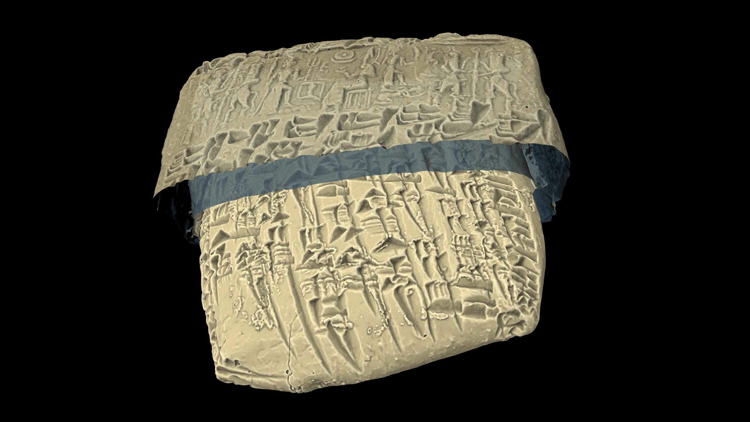

Unraveling ancient epistolary secrets (3rd place Participants' Choice Award)
Philipp Paetzold, Andreas Schropp, DESY, Andreas Beckert, Samaneh Ehteram, Stephan Olbrich, UHH & Cécile Michel, CNRS (France)
This visualization presents an X-ray micro-CT image of an Old Assyrian loan contract (ca. 1950–1850 BC, Louvre AO8295) found at Kanesh (modern Kültepe), captured at the Musée du Louvre, Paris, with the ENCI (Encrypting Non-destructively Cuneiform Inscriptions) system, a custom-built mobile X-ray micro-CT setup. This imaging was part of a project aimed at studying ancient cuneiform texts, especially those ancient sealed legal documents and enveloped letters whose contents have remained concealed within clay envelopes for over four millennia. What sets this image apart is the collaborative effort of an interdisciplinary team of engineers, physicists, computer scientists, and assyriologists, who successfully scanned, reconstructed, visualized, and deciphered the hidden text on site at the Louvre in under an hour, offering a non-destructive glimpse into the content of the 4000-year-old letter. Through such images, scholars gain insights into the societal life reflected in private letters and legal texts written in cuneiform on clay tablets by the Sumerians and Assyrians, used for daily communication between 5000 and 3000 years ago. The unveiled texts provide a unique perspective into ancient Mesopotamian life, drawing astonishing parallels to our own modern existence.

Cellular lightsaber fight (2nd place Public Choice Award)
Johanna Rettenmeier, Kendra Maass, Tatjana Wedig, Kristian Pajtler, DKFZ
To investigate communication processes in brain tumors, these brain tumor cells were genetically labeled with green and red membrane markers. Imaging with confocal fluorescent microscopy allows us to get detailed insights into their complex membranous connections. This particular image stands out for capturing an intense interaction between brain tumor cells, highlighting the complexity of cellular communication, reminiscent of handling a lightsaber. Scientifically, delving into the membrane dynamics of brain tumor cells offers insights into their complex communication strategies important for tumor growth and therapy resistance. This paves the way for innovative treatment approaches targeting membranous communication pathways.

Sugar and amino acid eaters
Yu-Le Wu, Tara Ziegelbauer, DKFZ
In miniaturized liver tumors, or liver organoids, proteins were labeled with antibodies carrying metal isotopes. The abundance of these isotopes, indicative of protein levels, was measured pixel by pixel using Multiplexed Ion Beam Imaging (MIBI). This technique specifically visualized various components within the cells: glucose and amino acid transporters in cyan and magenta, mitochondria in yellow as the cellular power plants, and cell nuclei in blue. This approach was undertaken with the goal of unraveling the connections between cellular metabolism and the organization of biological structures within patient-derived liver organoids. What sets this image apart is the observation that cells originating from the same tumor exhibit different nutritional intake preferences, indicated by cyan or magenta coloring. It appears that clusters of cells with identical preferences tend to be closely related, suggesting sibling relationships among them. Through such detailed imaging, the aim is to identify intracellular patterns that can predict how cancer cells will respond to various drugs, potentially guiding medical treatment strategies.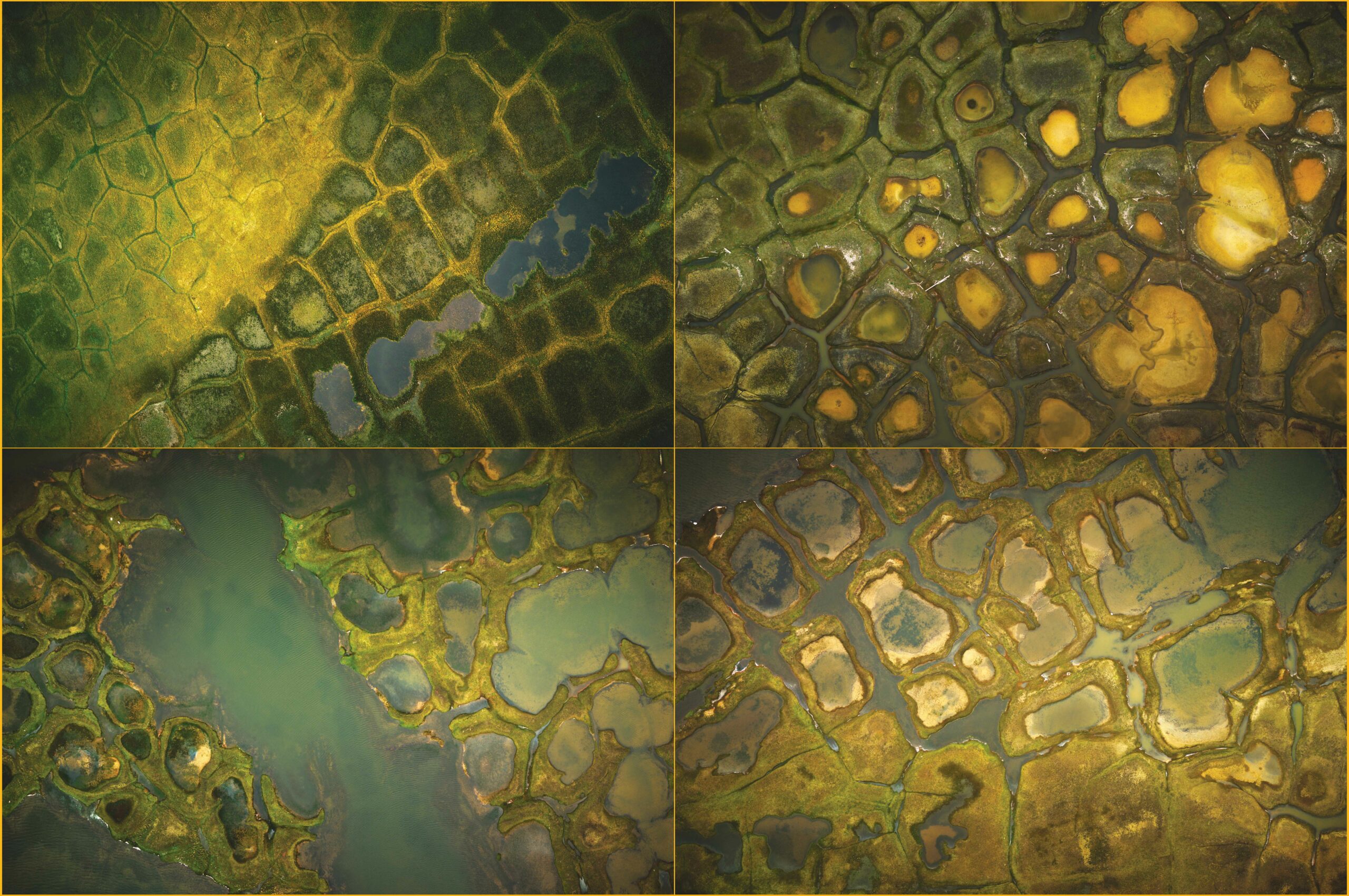

Collapsing arctic permafrost landscapes
Tilman Bucher, DLR & Guido Grosse, AWI
The image is a compilation of four individual shots taken by the DLR MACS aerial camera system aboard AWI's Polar-6 aircraft over the Alaskan Ikpikpuk River delta. With natural colors (RGB) enhanced for contrast and saturation, it offers a vivid portrayal of the area. These images are instrumental in monitoring the rapid thaw of permafrost, exacerbated by increasing saltwater flooding, to understand the processes of degradation affecting the region. This particular image stands out for its depiction of the evolutionary cycle of patterned ground degradation, showcasing the striking beauty and vulnerability of this wild environment amid signs of change. Through repeated surveys, these aerial images enable the identification and monitoring of permafrost degradation processes, determining rates of change. This data is crucial for training AI algorithms to interpret satellite data for global permafrost monitoring. Captured from a flight altitude of 500m, the images boast a ground resolution of 5 cm, offering detailed insights into the landscape's changing dynamics.

"Iron" maiden
Paul H. Kamm, HZB
This 3D-rendered tomographic image captures a brief moment in the dynamic growth of crystals within solidifying molten metal. The colored structures illustrate the intermetallic phases that form during the solidification of the AlNi alloy. These structures grow dendritically towards the center of the droplet, reminiscent of the medieval torture instrument of the iron maiden. The microstructure that forms plays a significant role in determining the material’s subsequent properties. Thus, understanding its development in relation to the production’s process parameters is of great importance. This 3D volume is not only recorded in a short time of 20 milliseconds with a sufficient resolution of a few micrometers over series of several seconds, but it is also three-, or possibly four-dimensional, unlike previous, comparable experiments. The technique of tomoscopy used for this image is suitable for providing insight into a variety of dynamic processes in different scientific disciplines. However, the immense volume of data generated requires significant effort in data analysis, where novel methods such as machine learning can offer substantial assistance.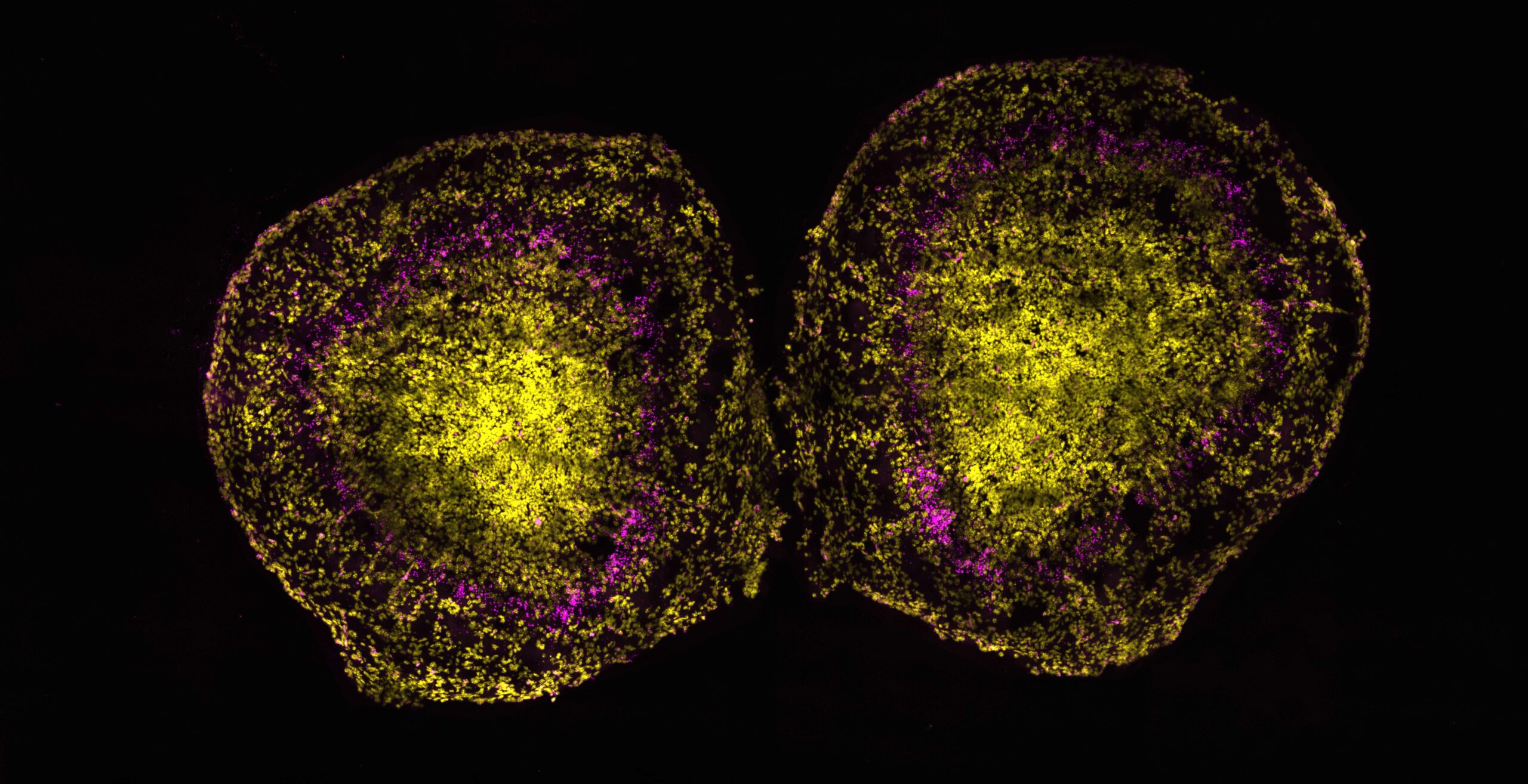

Pressure-sensing neurons in olfactory bulbs
Athanasios Balomenos, MDC
The image was captured using an Olympus IX83 and CSU-W1 spinning disk confocal system. The mRNA of interest was highlighted with fluorophores through RNAScope, and the final touches were added in ImageJ. This particular visual was obtained from sections of developing olfactory bulbs of postnatal mice, barely a day old, to visualize the RNA expression of the mechanosensitive channel Piezo2 in magenta, with cell nuclei marked in yellow. This image stands out for its ring-like pattern of Piezo2 expression in the two mirror-symmetric olfactory bulbs, prompting intriguing questions about why nature opts for such a unique pattern to express a pressure-sensitive protein in the brain's smell center. The scientific journey with this image led to the identification of specific neurons in the olfactory system that express Piezo2. For the first time, after culturing these cells, it was demonstrated that central olfactory neurons are capable of sensing mechanical stimuli, suggesting that the act of sniffing could activate these neurons.

Protein plymers targeting artificial vesicles
Marius Weismehl, MDC
This false-colored negative-stained transmission electron micrograph showcases protein-coated membranes at an impressive 73,000x nominal magnification, captured using a transmission electron microscope operating at 120 kV. The purpose of acquiring this image was to delve into the molecular mechanism of guanylate-binding proteins (GBPs), which play a critical role in host defense against bacterial pathogens by forming an antimicrobial protein coat on bacteria. What sets this image apart is its detailed capture of multiple binding events where protein polymers attach to artificial vesicles. These vesicles serve as a simplified model system, mimicking the bacterial surface, and illustrate the formation of a dense protein coat. The micrograph provides vital mechanistic insights into the assembly pathway of antimicrobial GBP1 at a high structural resolution. It aids in the understanding of GBP-orchestrated host immunity against bacterial infection.